• 文献检索
  • 文档翻译
  • 深度研究
  • 学术资讯
  • Suppr Zotero 插件Zotero 插件
  • 邀请有礼
  • 套餐&价格
  • 历史记录
应用&插件
Suppr Zotero 插件Zotero 插件浏览器插件Mac 客户端Windows 客户端微信小程序
定价
高级版会员购买积分包购买API积分包
服务
文献检索文档翻译深度研究API 文档MCP 服务
关于我们
关于 Suppr公司介绍联系我们用户协议隐私条款
关注我们

Suppr 超能文献

核心技术专利:CN118964589B侵权必究
粤ICP备2023148730 号-1Suppr @ 2026

文献检索

告别复杂PubMed语法,用中文像聊天一样搜索,搜遍4000万医学文献。AI智能推荐,让科研检索更轻松。

立即免费搜索

文件翻译

保留排版,准确专业,支持PDF/Word/PPT等文件格式,支持 12+语言互译。

免费翻译文档

深度研究

AI帮你快速写综述,25分钟生成高质量综述,智能提取关键信息,辅助科研写作。

立即免费体验

通过免疫细胞化学方法显示,促黄体素释放激素(LHRH)纤维有丰富的室管膜覆盖,其图像类似高尔基染色的图像。

Rich ependymal investment of luliberin (LHRH) fibers revealed immunocytochemically in an image like that from Golgi stain.

作者信息

Burchanowski B J, Knigge K M, Sternberger L A

出版信息

Proc Natl Acad Sci U S A. 1979 Dec;76(12):6671-4. doi: 10.1073/pnas.76.12.6671.

DOI:10.1073/pnas.76.12.6671
PMID:392523
原文链接:https://pmc.ncbi.nlm.nih.gov/articles/PMC411930/
Abstract

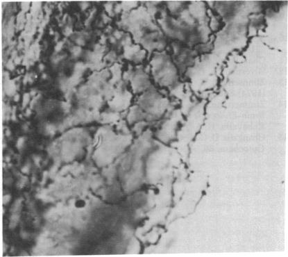
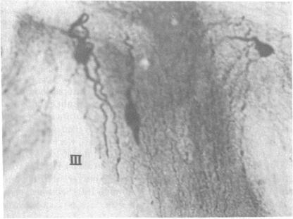
Immunospecific staining in 100-micron-thick Vibratome section by the unlabeled antibody method resembles Golgi staining and reveals an abundance of luliberin- (luteinizing hormone releasing hormone, LHRH) positive cells and fibers in close contact with the surface of the third ventricle. The polarity of LHRH cells can be seen and processes can be traced for several millimeters. In the medial preoptic and suprachiasmatic areas bipolar LHRH neurons send short stout processes to the ventricular surface and long processes toward the organum vasculosum laminae terminalis. These cells resemble the receptor cells contacting the cerebrospinal fluid that have been described by Vigh and Vigh-Teichmann [Vigh, B. & Vigh-Teichmann, I. (1973) Int. Rev. Cytol. 35, 189-251]. In the septal region some bipolar neurons send both of their processes towards the ventricular surface. LHRH neurons in the nucleus of the anterior commissure and the bed nucleus of the stria terminalis project over the anterior commissure to form a dense plexus of fibers in the subfornical organ. The proximity of LHRH perikarya and fibers to the ventricular surface supports the hypothesis [Knigge, K.M., Joseph, S.A., Scott, D.E. & Jacobs, J.J. (1971) in The Neuroendocrinology of Human Reproduction, eds. Mack, H.C. & Sherman, A.J., (Thomas, Springfield, IL), pp. 6-22.] that the cerebrospinal fluid functions in neuroendocrine control mechanisms.

摘要

通过未标记抗体法对100微米厚的振动切片机切片进行免疫特异性染色,类似于高尔基染色,显示出大量与第三脑室表面紧密接触的促黄体素释放激素(LHRH)阳性细胞和纤维。可以看到LHRH细胞的极性,其突起可追踪数毫米。在内侧视前区和视交叉上区,双极LHRH神经元向脑室表面发出短而粗的突起,向终板血管器发出长突起。这些细胞类似于Vigh和Vigh-Teichmann [Vigh, B. & Vigh-Teichmann, I. (1973) Int. Rev. Cytol. 35, 189 - 251]所描述的与脑脊液接触的受体细胞。在隔区,一些双极神经元将其两个突起都伸向脑室表面。前连合核和终纹床核中的LHRH神经元向前连合投射,在穹窿下器官形成密集的纤维丛。LHRH核周体和纤维靠近脑室表面支持了这样一种假说[Knigge, K.M., Joseph, S.A., Scott, D.E. & Jacobs, J.J. (1971) in The Neuroendocrinology of Human Reproduction, eds. Mack, H.C. & Sherman, A.J., (Thomas, Springfield, IL), pp. 6 - 22.],即脑脊液在神经内分泌控制机制中起作用。

https://cdn.ncbi.nlm.nih.gov/pmc/blobs/e98d/411930/b32c347650ae/pnas00012-0657-b.jpg
https://cdn.ncbi.nlm.nih.gov/pmc/blobs/e98d/411930/1561b9077366/pnas00012-0656-a.jpg
https://cdn.ncbi.nlm.nih.gov/pmc/blobs/e98d/411930/e55266fe145d/pnas00012-0656-b.jpg
https://cdn.ncbi.nlm.nih.gov/pmc/blobs/e98d/411930/763dbe6d566e/pnas00012-0656-c.jpg
https://cdn.ncbi.nlm.nih.gov/pmc/blobs/e98d/411930/a63371cf0f6e/pnas00012-0656-d.jpg
https://cdn.ncbi.nlm.nih.gov/pmc/blobs/e98d/411930/ab18c4adbdb1/pnas00012-0657-a.jpg
https://cdn.ncbi.nlm.nih.gov/pmc/blobs/e98d/411930/b32c347650ae/pnas00012-0657-b.jpg

相似文献

1
Rich ependymal investment of luliberin (LHRH) fibers revealed immunocytochemically in an image like that from Golgi stain.通过免疫细胞化学方法显示,促黄体素释放激素(LHRH)纤维有丰富的室管膜覆盖,其图像类似高尔基染色的图像。
Proc Natl Acad Sci U S A. 1979 Dec;76(12):6671-4. doi: 10.1073/pnas.76.12.6671.
2
Improved visualization of luteinizing hormone releasing hormone pathways by immunocytochemical staining of thick vibratome sections.
J Histochem Cytochem. 1980 Apr;28(4):361-3. doi: 10.1177/28.4.6989900.
3
Immunocytochemical distribution of LHRH neurons and processes in the rat: hypothalamic and extrahypothalamic locations.大鼠中促黄体生成素释放激素(LHRH)神经元及突起的免疫细胞化学分布:下丘脑及下丘脑外定位
Cell Tissue Res. 1982;221(3):493-504. doi: 10.1007/BF00215698.
4
LHRH immunopositive cells and their projections to the median eminence and organum vasculosum of the lamina terminalis.促性腺激素释放激素免疫阳性细胞及其向正中隆起和终板血管器的投射。
J Comp Neurol. 1982 Aug 10;209(3):287-300. doi: 10.1002/cne.902090307.
5
The luteinizing hormone-releasing hormone (LHRH) systems in the rat brain.大鼠大脑中的促黄体生成激素释放激素(LHRH)系统。
Neuroendocrinology. 1982 Dec;35(6):429-38. doi: 10.1159/000123419.
6
Immunocytochemical demonstration of neuropeptide Y and luteinizing hormone-releasing hormone-immunoreactive structures in the organum vasculosum laminae terminalis of juvenile gilts.幼年母猪终板血管器中神经肽Y和促黄体生成素释放激素免疫反应性结构的免疫细胞化学证明
Neurosci Lett. 1993 Aug 6;158(1):21-4. doi: 10.1016/0304-3940(93)90602-h.
7
Immunocytochemical localization of luteinizing hormone-releasing hormone (LHRH) pathways in the sheep brain during anestrus and the mid-luteal phase of the estrous cycle.发情周期乏情期和黄体中期绵羊大脑中促黄体生成激素释放激素(LHRH)通路的免疫细胞化学定位。
J Comp Neurol. 1986 Feb 1;244(1):19-35. doi: 10.1002/cne.902440103.
8
The distribution of luteinizing hormone-releasing hormone (LHRH) neurons and fibers in the Formosan rock-monkey.黄体生成素释放激素(LHRH)神经元和纤维在台湾猕猴中的分布。
Proc Natl Sci Counc Repub China B. 1988 Jul;12(3):156-62.
9
Ependymoneuronal specializations between LHRH fibers and cells of the cerebroventricular system.促性腺激素释放激素(LHRH)纤维与脑室系统细胞之间的室管膜神经元特化
Cell Tissue Res. 1985;242(2):301-11. doi: 10.1007/BF00214542.
10
Localization of luteinizing hormone-releasing hormone in the forebrain of the pig.促黄体生成素释放激素在猪前脑的定位
Biol Reprod. 1988 Oct;39(3):665-72. doi: 10.1095/biolreprod39.3.665.

引用本文的文献

1
Highlights of neuroanatomical discoveries of the mammalian gonadotropin-releasing hormone system.哺乳动物促性腺激素释放激素系统神经解剖学发现要点。
J Neuroendocrinol. 2022 May;34(5):e13115. doi: 10.1111/jne.13115. Epub 2022 May 3.
2
A novel protocol of whole mount electro-immunofluorescence staining.一种新型的全组织电免疫荧光染色方案。
Mol Vis. 2009;15:505-17. Epub 2009 Mar 6.
3
Immunocytochemical distribution of LHRH neurons and processes in the rat: hypothalamic and extrahypothalamic locations.大鼠中促黄体生成素释放激素(LHRH)神经元及突起的免疫细胞化学分布:下丘脑及下丘脑外定位

本文引用的文献

1
Comparative ultrastructure of the cerebrospinal fluid-contacting neurons.脑脊液接触神经元的超微结构比较
Int Rev Cytol. 1973;35:189-251. doi: 10.1016/s0074-7696(08)60355-1.
2
Distribution of indolealkylamine nerve terminals in the ventricles of the rat brain.吲哚烷基胺神经末梢在大鼠脑室中的分布。
Z Zellforsch Mikrosk Anat. 1973 Nov 15;144(4):511-22. doi: 10.1007/BF00307377.
3
Localization of gonadotropin-releasing hormone (Gn-RH) in the hypothalamus of the mouse by immunoperoxidase technique.采用免疫过氧化物酶技术对小鼠下丘脑促性腺激素释放激素(Gn-RH)进行定位。
Cell Tissue Res. 1982;221(3):493-504. doi: 10.1007/BF00215698.
4
Effects of monosodium glutamate on the development of intraventricular axons in the rat hypothalamus.谷氨酸钠对大鼠下丘脑室内轴突发育的影响。
Anat Embryol (Berl). 1983;168(3):433-44. doi: 10.1007/BF00304279.
5
Ependymoneuronal specializations between LHRH fibers and cells of the cerebroventricular system.促性腺激素释放激素(LHRH)纤维与脑室系统细胞之间的室管膜神经元特化
Cell Tissue Res. 1985;242(2):301-11. doi: 10.1007/BF00214542.
Endocrinology. 1974 Jul;95(1):1-8. doi: 10.1210/endo-95-1-1.
4
Modification of brain and CSF LH-RH following deafferentation.去传入神经后大脑和脑脊液中促性腺激素释放激素的改变。
Neuroendocrinology. 1975;18(1):131-5. doi: 10.1159/000122391.
5
Indoleamine neurons and their processes in the normal rat brain and in chronic diet-induced thiamine deficiency demonstrated by uptake of 3H-serotonin.通过3H-血清素摄取显示正常大鼠脑和慢性饮食诱导的硫胺素缺乏中的吲哚胺神经元及其突起。
J Comp Neurol. 1977 Dec 15;176(4):467-93. doi: 10.1002/cne.901760402.
6
The cerebrospinal fluid as a pathway in neuroendocrine integration.脑脊液作为神经内分泌整合的一条途径。
J Endocrinol. 1976 Dec;71(3):407-43. doi: 10.1677/joe.0.0710407.
7
Distribution of growth hormone-release-inhibiting hormone (somatostatin) in the rat brain as observed with immunocytochemistry.用免疫细胞化学法观察生长抑素(促生长素释放抑制激素)在大鼠脑中的分布。
Anat Rec. 1976 Nov;186(3):343-55. doi: 10.1002/ar.1091860302.
8
Distribution of luteinizing hormone-releasing hormone (LHRH) in the guinea pig brain.黄体生成素释放激素(LHRH)在豚鼠脑中的分布。
Endocrinology. 1976 Jul;99(1):30-41. doi: 10.1210/endo-99-1-30.
9
On the immunocytochemical localization of the vasoactive intestinal polypeptide.关于血管活性肠肽的免疫细胞化学定位
J Histochem Cytochem. 1979 May;27(5):936-8. doi: 10.1177/27.5.479555.
10
Cellular localization and ontogeny of immunoreactive vasoactive intestinal polypeptide (VIP) in the chicken gut.
Cell Tissue Res. 1979 Feb 15;196(2):193-201. doi: 10.1007/BF00240095.