Nallathambi Kowsalya, Elangovan Ramnath, J Selvakumar, Jayden Ebenezer, Maganti Dinesh C, Soman Dona
Department of Periodontics, Adhiparasakthi Dental College and Hospital, Melmaruvathur, IND.
Department of Periodontology, School of Dentistry, University of Rwanda, Kigali, RWA.
Cureus. 2024 Aug 16;16(8):e66975. doi: 10.7759/cureus.66975. eCollection 2024 Aug.
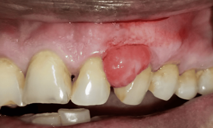
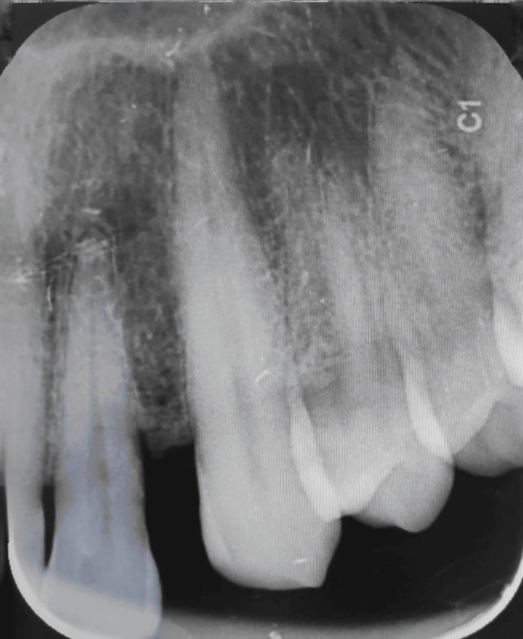
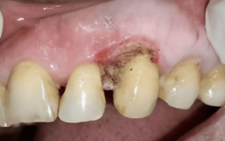
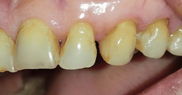
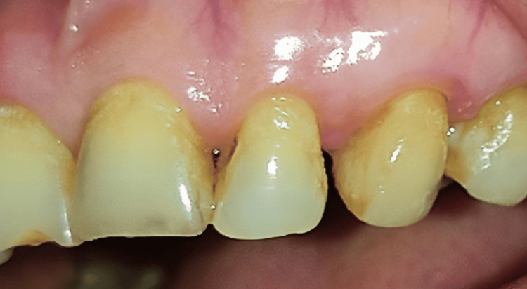
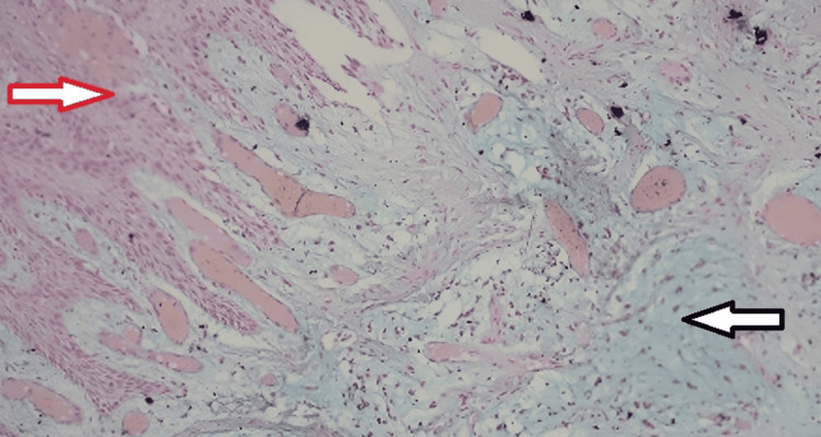
Myxomas are a group of benign tumors that have a common histologic appearance of fibrous and myxoid ground substance. According to literature, myxomas often occur between the ages of 30 and 50 years. Very often, intraoral soft tissue myxoma can be misinterpreted as malignant and is difficult to differentiate from other tumors with myxoid stroma. Of the different variants of soft tissue myxoma, intraoral is an extremely rare, slow-growing, benign ectomesenchymal tumor. We report a case of a 75-year-old female who presented with soft tissue swelling in the upper front tooth region. No history of pain over the lesion or bleeding was seen during brushing. On intraoral examination, a lesion measuring 2 x 3 cm was seen in the interdental papilla involving the attached gingiva of 22 and 23. An excisional biopsy of the lesion using a diode laser followed by low-level laser therapy revealed oral soft tissue fibromyxoma without odontogenic origin. A case of oral soft tissue myxoma is presented for its rarity and differential diagnosis of localized oral cavity lesions.
黏液瘤是一组良性肿瘤,具有纤维性和黏液样基质的共同组织学表现。根据文献记载,黏液瘤常发生于30至50岁之间。口腔软组织黏液瘤常常被误诊为恶性肿瘤,并且很难与其他具有黏液样基质的肿瘤相鉴别。在软组织黏液瘤的不同变体中,口腔内黏液瘤是一种极其罕见、生长缓慢的良性外间充质肿瘤。我们报告一例75岁女性患者,其上前牙区出现软组织肿胀。病变部位无疼痛史,刷牙时也未见出血。口腔检查发现,在22和23牙间乳头处有一个2×3厘米大小的病变,累及附着龈。使用二极管激光对病变进行切除活检,随后进行低强度激光治疗,结果显示为非牙源性的口腔软组织纤维黏液瘤。本文报告一例口腔软组织黏液瘤,因其罕见性以及对局限性口腔病变的鉴别诊断意义而被呈现。