Iwao Yuma, Kawata Naoko, Sekiguchi Yuki, Haneishi Hideaki
Center for Frontier Medical Engineering, Chiba University, 1-33, Yayoi-cho, Inage-ku, Chiba-shi, Chiba, 263-8522, Japan.
Institute for Quantum Medical Science, National Institutes for Quantum Science and Technology, 4-9-1, Anagawa, Inage-ku, Chiba-shi, Chiba, 263-8555, Japan.
Heliyon. 2024 Aug 31;10(17):e37272. doi: 10.1016/j.heliyon.2024.e37272. eCollection 2024 Sep 15.
To analyze morphological changes in patients with COVID-19-associated pneumonia over time, a nonrigid registration technique is required that reduces differences in respiratory phase and imaging position and does not excessively deform the lesion region. A nonrigid registration method using deep learning was applied for lung field alignment, and its practicality was verified through quantitative evaluation, such as image similarity of whole lung region and image similarity of lesion region, as well as visual evaluation by a physician.
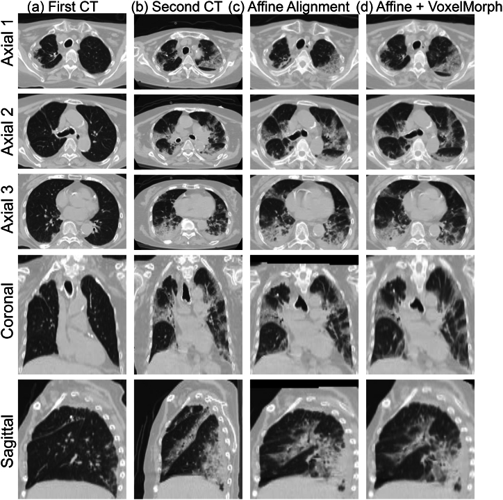
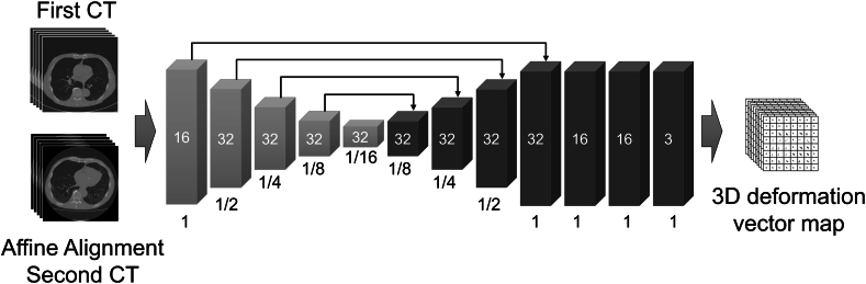
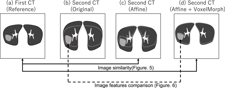
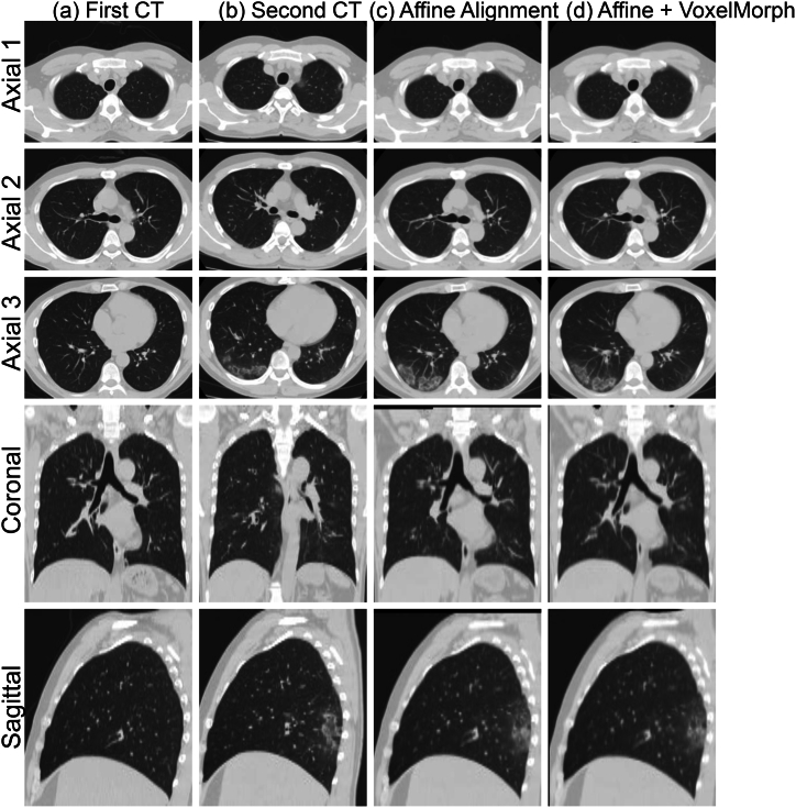
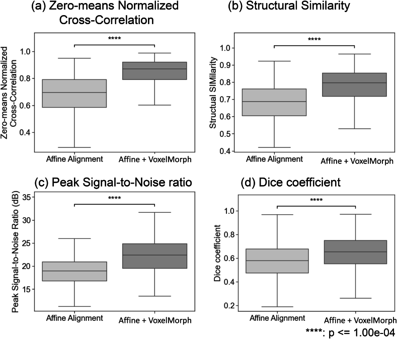
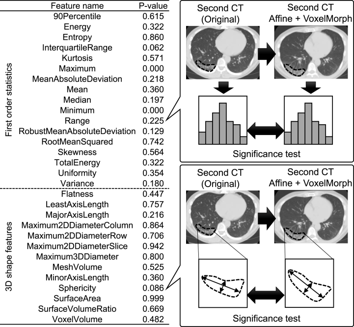
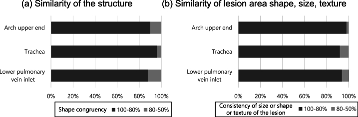
First, the lung field positions and sizes of the first and second CT images were roughly matched using a classical registration method based on iterative calculations as a preprocessing step. Then, voxel-by-voxel transformation was performed using VoxelMorph, a nonrigid deep learning registration method. As an objective evaluation, the similarity of the images was calculated. To evaluate the invariance of image features in the lesion site, primary statistics and 3D shape features were calculated and statistically analyzed. Furthermore, as a subjective evaluation, the similarity of images and whether nonrigid transformation caused unnatural changes in the shape and size of the lesion region were visually evaluated by a pulmonologist.
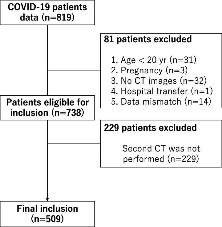
The proposed method was applied to 509 patient data points with high image similarity. The variances in histogram characteristics before and after image deformation were confirmed. Visual evaluation confirmed the agreement between the shape and internal structure of the lung field and the natural deformation of the lesion region.
The developed nonrigid registration method was shown to be effective for quantitative time series analysis of the lungs.
为了分析新型冠状病毒肺炎相关肺炎患者随时间的形态变化,需要一种非刚性配准技术,该技术可减少呼吸相位和成像位置的差异,且不会使病变区域过度变形。将一种使用深度学习的非刚性配准方法应用于肺野对齐,并通过全肺区域图像相似度、病变区域图像相似度等定量评估以及医生的视觉评估来验证其实用性。
首先,作为预处理步骤,使用基于迭代计算的经典配准方法大致匹配第一幅和第二幅CT图像的肺野位置和大小。然后,使用非刚性深度学习配准方法VoxelMorph进行逐体素变换。作为客观评估,计算图像的相似度。为了评估病变部位图像特征的不变性,计算并统计分析主要统计量和三维形状特征。此外,作为主观评估,由肺科医生对图像的相似度以及非刚性变换是否导致病变区域的形状和大小出现不自然变化进行视觉评估。
所提出的方法应用于509个具有高图像相似度的患者数据点。确认了图像变形前后直方图特征的差异。视觉评估证实了肺野形状和内部结构与病变区域自然变形之间的一致性。
所开发的非刚性配准方法被证明对肺部的定量时间序列分析有效。