Sharma Swati, Shamjetsabam Nandibala Devi, Chauhan Kirti, Yashavarddhan M H, Gautam Poonam, Prakash Prem, Choudhary Priyanka, Chhabra Satnam S, Acharya Rajesh, Kalra Samir K, Gupta Anshul, Jain Sunila, Ganguly Nirmal K, Rana Rashmi
Department of Biotechnology and Research, Sir Ganga Ram Hospital.
Central Proteomics Facility, ICMR-National Institute of Pathology.
Int J Surg. 2024 Dec 1;110(12):7434-7446. doi: 10.1097/JS9.0000000000002054.
Meningioma is the most prevalent primary intracranial brain tumor and accounts for one-third of all CNS tumors. Meningioma is known to be the most common yet life-threatening brain tumor with a higher recurrence rate. Globally, there is an increase in the healthcare burden due to meningioma and hence in its research. The present clinical approach includes surgical resection, chemotherapy, and radiation therapies to which the malignancy does not seem to respond efficiently. Targeted therapies and molecular markers provide elite patient treatment and care for individuals suffering from meningiomas as compared to conventional measures. Although there is proteomic data on meningioma the knowledge of potential biomarkers differentiating the grades is scarce. To identify the best set of biomarkers, validation of reported markers in large and independent sample cohorts in the future is necessary.
A total of 12 samples, 3 each of control (which made pool 1) meningioma grade I (which made 2 sets: pool 2 and pool 3), and meningioma grade II (which made pool 4) were taken for LC-MS/MS. After this, the expression of three proteins was checked by immunocytochemistry, flow cytometry, and western blotting.
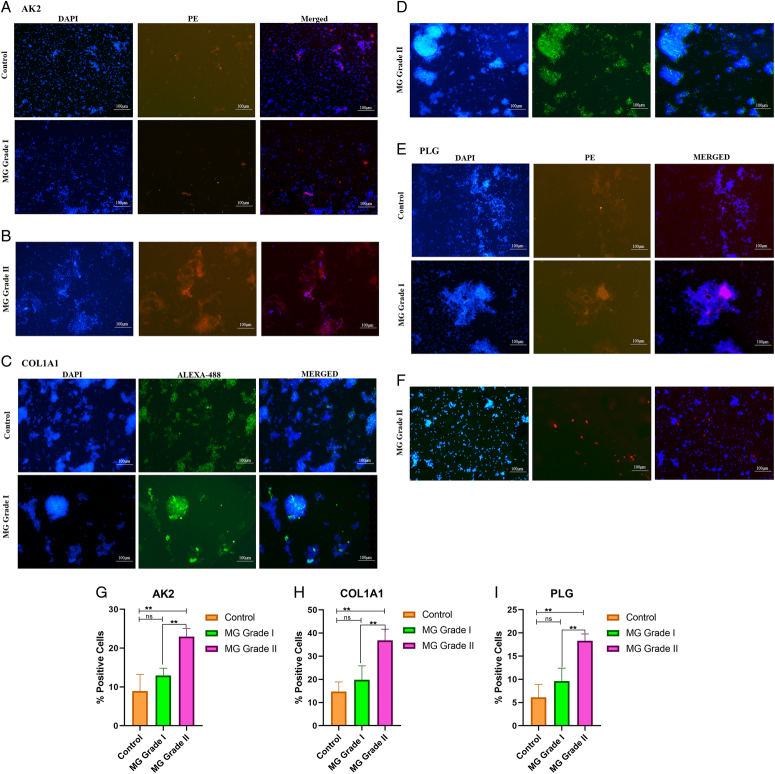
Protein expression was analyzed using various techniques like mass spectrometry, immunocytochemistry, flow cytometry, and western blotting. Mass spectrometry is the most commonly used standard and reliable technique for identifying and quantifying protein expression. We got three highly upregulated proteins namely AK2, COL1A1, and PLG using this technique. The biomarker potential of these proteins was further checked by ICC, western blotting, and flow cytometry. Three important proteins were found to be upregulated namely, AK2 (Adenylate kinase 2), COL1A1 (collagen 1A1), and PLG (plasminogen). The order of increased protein expression was control < MG grade I < MG grade II according to mass spectrometry and western blotting. In immunocytochemistry, we found that COL1A1 expression increases significantly with grades in comparison to control. Similarly, AK2 and PLG also showed little increase but not as much as COL1A1. In flow cytometry, PLG showed higher upregulation in grades than control. While AK2 and COL1A1 showed little increase in expression in grades than control. All techniques, especially mass spectrometry and western blotting, presented higher expression of these proteins in grades as compared to control.
In the quest to find a suitable therapeutic marker, this study incorporates quantitative labeling and detection followed by flow cytometry, immunocytochemistry, and western blotting for early diagnosis and treatment of meningioma. The article further explores the efficacy of some proteins namely AK2, COL1A1, and PLG to be the targeted molecules.
脑膜瘤是最常见的原发性颅内脑肿瘤,占所有中枢神经系统肿瘤的三分之一。脑膜瘤是已知最常见但危及生命且复发率较高的脑肿瘤。在全球范围内,由于脑膜瘤导致的医疗负担增加,因此其研究也在增加。目前的临床方法包括手术切除、化疗和放疗,但恶性肿瘤对这些方法似乎反应不佳。与传统措施相比,靶向治疗和分子标志物为患有脑膜瘤的个体提供了优质的患者治疗和护理。尽管有关于脑膜瘤的蛋白质组学数据,但区分不同级别的潜在生物标志物的知识却很匮乏。为了确定最佳的生物标志物组合,未来有必要在大型独立样本队列中对已报道的标志物进行验证。
共采集了12个样本,其中对照组3个(组成样本池1),I级脑膜瘤6个(组成2个样本池:样本池2和样本池3),II级脑膜瘤3个(组成样本池4)用于液相色谱-串联质谱分析(LC-MS/MS)。此后,通过免疫细胞化学、流式细胞术和蛋白质免疫印迹法检测三种蛋白质的表达。
使用质谱分析、免疫细胞化学、流式细胞术和蛋白质免疫印迹法等多种技术分析蛋白质表达。质谱分析是用于鉴定和定量蛋白质表达的最常用的标准且可靠的技术。使用该技术我们得到了三种高度上调的蛋白质,即腺苷酸激酶2(AK2)、I型胶原蛋白α1链(COL1A1)和纤溶酶原(PLG)。通过免疫细胞化学、蛋白质免疫印迹法和流式细胞术进一步检测了这些蛋白质的生物标志物潜力。发现三种重要蛋白质上调,即AK2(腺苷酸激酶2)、COL1A1(I型胶原蛋白α1链)和PLG(纤溶酶原)。根据质谱分析和蛋白质免疫印迹法,蛋白质表达增加的顺序为对照组<I级脑膜瘤<II级脑膜瘤。在免疫细胞化学中,我们发现与对照组相比,COL1A1的表达随级别显著增加。同样,AK2和PLG也有少量增加,但不如COL1A1增加得多。在流式细胞术中,PLG在不同级别中的上调程度高于对照组。而AK2和COL1A1在不同级别中的表达增加幅度小于对照组。所有技术,尤其是质谱分析和蛋白质免疫印迹法,均显示这些蛋白质在不同级别中的表达高于对照组。
为了寻找合适的治疗标志物,本研究采用定量标记和检测,随后进行流式细胞术、免疫细胞化学和蛋白质免疫印迹法,以实现脑膜瘤的早期诊断和治疗。本文进一步探讨了一些蛋白质,即AK2、COL1A1和PLG作为靶向分子的有效性。