Wallace H. Coulter Department of Biomedical Engineering, Atlanta, GA, USA.
Parker H. Petit Institute for Bioengineering and Biosciences, Atlanta, GA, USA.
Nat Commun. 2024 Sep 27;15(1):8339. doi: 10.1038/s41467-024-52565-2.
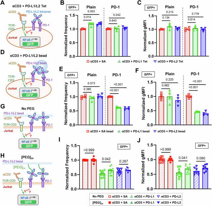
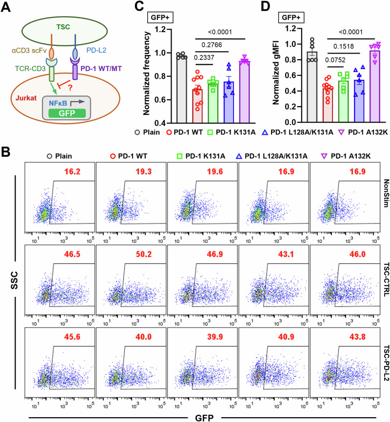
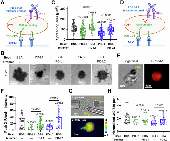
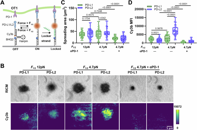
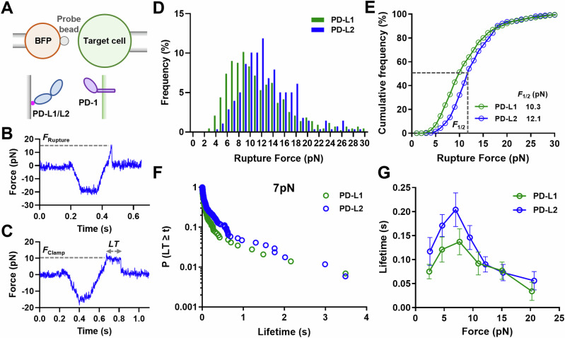
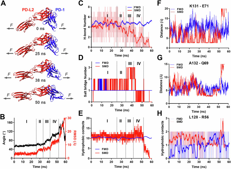
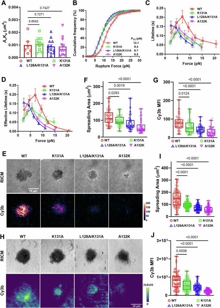
Despite the success of PD-1 blockade in cancer therapy, how PD-1 initiates signaling remains unclear. Soluble PD-L1 is found in patient sera and can bind PD-1 but fails to suppress T cell function. Here, we show that PD-1 function is reduced when mechanical support on ligand is removed. Mechanistically, cells exert forces to PD-1 and prolong bond lifetime at forces <7 pN (catch bond) while accelerate dissociation at forces >8pN (slip bond). Molecular dynamics of PD-1-PD-L2 complex suggests force may cause relative rotation and translation between the two molecules yielding distinct atomic contacts not observed in the crystal structure. Compared to wild-type, PD-1 mutants targeting the force-induced distinct interactions maintain the same binding affinity but suppressed/eliminated catch bond, lowered rupture force, and reduced inhibitory function. Our results uncover a mechanism for cells to probe the mechanical support of PD-1-PD-Ligand bonds using endogenous forces to regulate PD-1 signaling.
尽管 PD-1 阻断在癌症治疗中取得了成功,但 PD-1 如何引发信号仍不清楚。可溶性 PD-L1 存在于患者血清中,可与 PD-1 结合,但不能抑制 T 细胞功能。在这里,我们表明,当配体的机械支撑被去除时,PD-1 的功能会降低。从机制上讲,当力<7 pN 时,细胞对 PD-1 施加力并延长键的寿命(捕捉键),而当力>8 pN 时,加速键的解离(滑动键)。PD-1-PD-L2 复合物的分子动力学表明,力可能导致两个分子之间的相对旋转和平移,产生在晶体结构中观察不到的独特原子接触。与野生型相比,针对力诱导的独特相互作用的 PD-1 突变体保持相同的结合亲和力,但抑制/消除了捕捉键,降低了断裂力,并降低了抑制功能。我们的研究结果揭示了细胞利用内源性力探测 PD-1-PD-Ligand 键的机械支撑的机制,从而调节 PD-1 信号。