From the ‡Department of Biochemistry, Vanderbilt University School of Medicine, Nashville, Tennessee.
§Hematology/Oncology Division, Department of Medicine, Vanderbilt University School of Medicine, Nashville, Tennessee 37232.
Mol Cell Proteomics. 2017 Oct;16(10):1705-1717. doi: 10.1074/mcp.RA117.000037. Epub 2017 May 25.
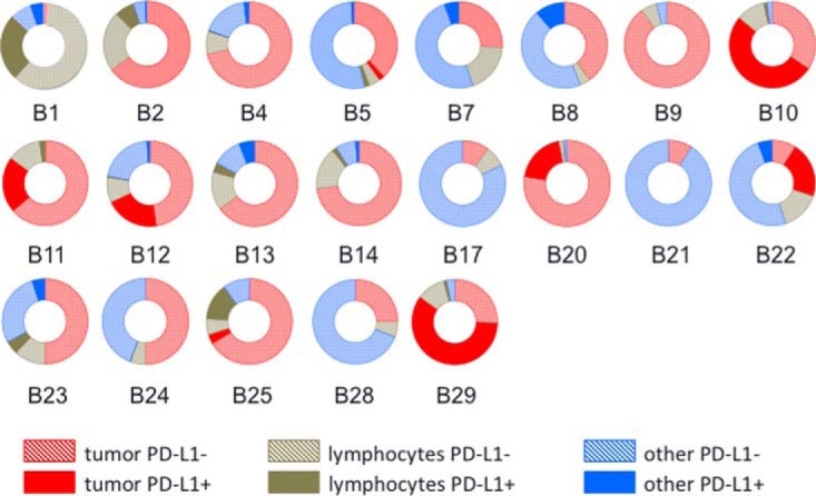
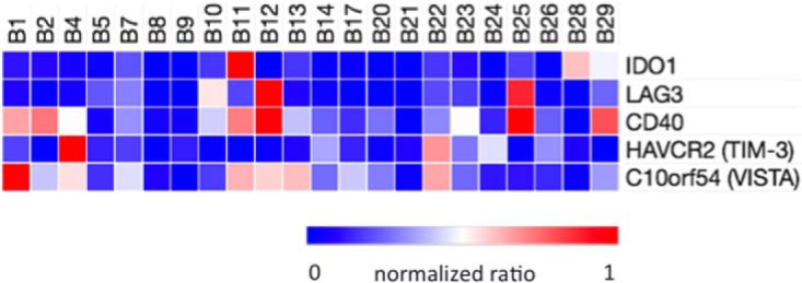
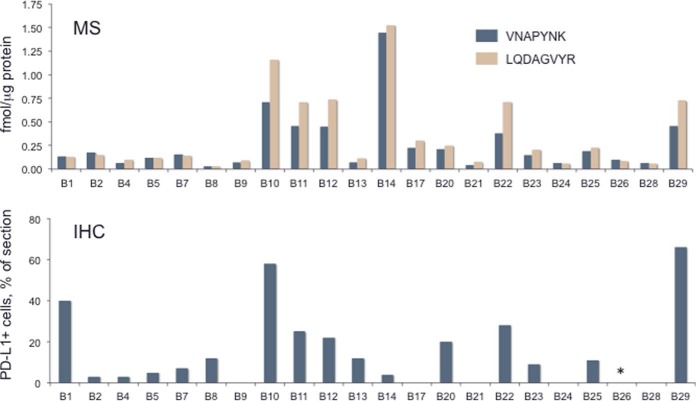
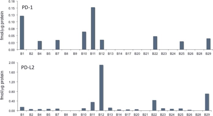
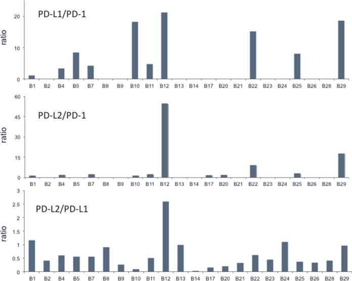
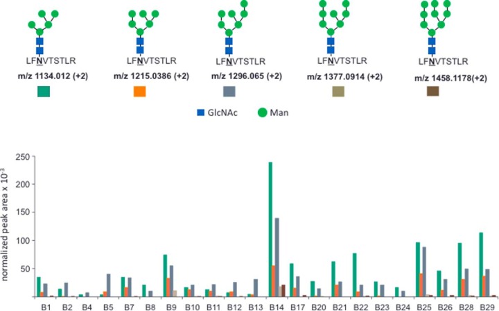
Quantitative assessment of key proteins that control the tumor-immune interface is one of the most formidable analytical challenges in immunotherapeutics. We developed a targeted MS platform to quantify programmed cell death-1 (PD-1), programmed cell death 1 ligand 1 (PD-L1), and programmed cell death 1 ligand 2 (PD-L2) at fmol/microgram protein levels in formalin fixed, paraffin-embedded sections from 22 human melanomas. PD-L1 abundance ranged 50-fold, from ∼0.03 to 1.5 fmol/microgram protein and the parallel reaction monitoring (PRM) data were largely concordant with total PD-L1-positive cell content, as analyzed by immunohistochemistry (IHC) with the E1L3N antibody. PD-1 was measured at levels up to 20-fold lower than PD-L1, but the abundances were not significantly correlated (r = 0.062, = 0.264). PD-1 abundance was weakly correlated (r = 0.3057, = 0.009) with the fraction of lymphocytes and histiocytes in sections. PD-L2 was measured from 0.03 to 1.90 fmol/microgram protein and the ratio of PD-L2 to PD-L1 abundance ranged from 0.03 to 2.58. In 10 samples, PD-L2 was present at more than half the level of PD-L1, which suggests that PD-L2, a higher affinity PD-1 ligand, is sufficiently abundant to contribute to T-cell downregulation. We also identified five branched mannose and N-acetylglucosamine glycans at PD-L1 position N192 in all 22 samples. Extent of PD-L1 glycan modification varied by ∼10-fold and the melanoma with the highest PD-L1 protein abundance and most abundant glycan modification yielded a very low PD-L1 IHC estimate, thus suggesting that N-glycosylation may affect IHC measurement and PD-L1 function. Additional PRM analyses quantified immune checkpoint/co-regulator proteins LAG3, IDO1, TIM-3, VISTA, and CD40, which all displayed distinct expression independent of PD-1, PD-L1, and PD-L2. Targeted MS can provide a next-generation analysis platform to advance cancer immuno-therapeutic research and diagnostics.
定量评估控制肿瘤免疫界面的关键蛋白是免疫治疗中最具挑战性的分析难题之一。我们开发了一种靶向 MS 平台,可在 22 个人黑色素瘤福尔马林固定石蜡包埋切片中以 fmol/μg 蛋白水平定量检测程序性细胞死亡蛋白 1 (PD-1)、程序性细胞死亡配体 1 (PD-L1)和程序性细胞死亡配体 2 (PD-L2)。PD-L1 的丰度范围为 50 倍,从约 0.03 到 1.5 fmol/μg 蛋白,平行反应监测 (PRM) 数据与免疫组化 (IHC) 用 E1L3N 抗体分析的总 PD-L1 阳性细胞含量基本一致。PD-1 的测量水平比 PD-L1 低 20 倍,但丰度无显著相关性 (r = 0.062, = 0.264)。PD-1 的丰度与切片中淋巴细胞和组织细胞的比例呈弱相关 (r = 0.3057, = 0.009)。PD-L2 的测量范围为 0.03 至 1.90 fmol/μg 蛋白,PD-L2 与 PD-L1 丰度的比值范围为 0.03 至 2.58。在 10 个样本中,PD-L2 的水平超过 PD-L1 的一半,这表明作为 PD-1 高亲和力配体的 PD-L2 含量丰富,足以抑制 T 细胞。我们还在所有 22 个样本中 PD-L1 的 N192 位置鉴定了五个分支的甘露糖和 N-乙酰葡萄糖胺聚糖。PD-L1 糖基化修饰的程度变化约 10 倍,PD-L1 蛋白丰度最高和糖基化修饰最丰富的黑色素瘤产生的 PD-L1 IHC 估计值非常低,这表明 N-糖基化可能会影响 IHC 测量和 PD-L1 功能。额外的 PRM 分析定量了免疫检查点/共调节剂蛋白 LAG3、IDO1、TIM-3、VISTA 和 CD40,它们的表达均与 PD-1、PD-L1 和 PD-L2 无关。靶向 MS 可以提供下一代分析平台,以推进癌症免疫治疗研究和诊断。