Kouznetsova Anna, Valentiniene Sonata, Liu Jian-Guo, Kitajima Tomoya S, Brismar Hjalmar, Höög Christer
Department of Cell and Molecular Biology, Karolinska Institutet, Stockholm, Sweden.
Laboratory for Chromosome Segregation, RIKEN Center for Biosystems Dynamics Research, Kobe, Japan.
Front Cell Dev Biol. 2024 Sep 17;12:1470981. doi: 10.3389/fcell.2024.1470981. eCollection 2024.
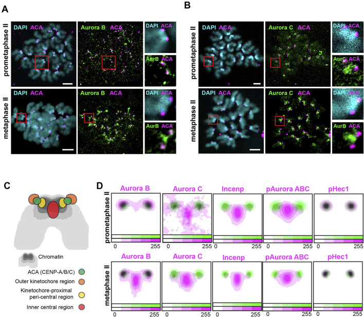
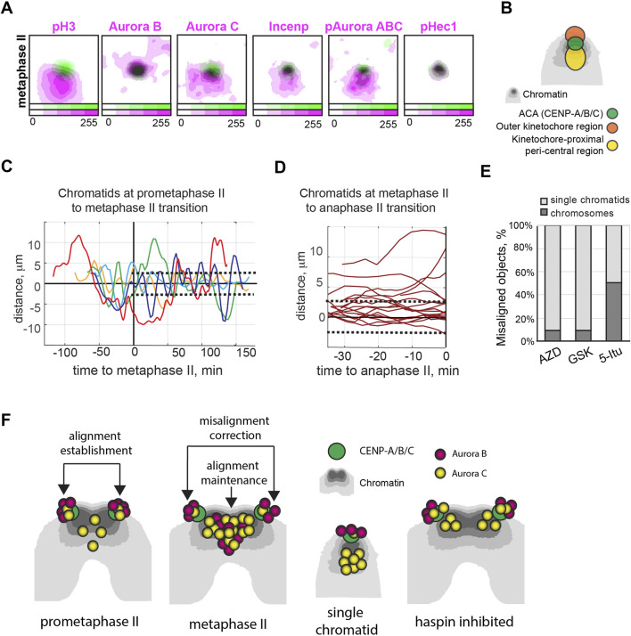
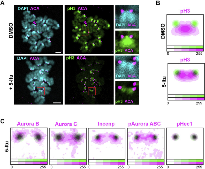
Correct chromosome segregation is essential to preserve genetic integrity. The two protein kinases, Aurora B and its meiotic homolog Aurora C, regulate attachments between chromosomal kinetochores and microtubules, thereby contributing to the accuracy of the chromosome segregation process. Here we performed a detailed examination of the localization and activity of Aurora B/C kinases, their partner Incenp and the kinetochore target Hec1, during the second meiotic division in mouse oocytes. We found that a majority of Aurora B and C changed their localization from the outer kinetochore region of chromosomes at prometaphase II to an inner central region localized between sister centromeres at metaphase II. Depletion of the Aurora B/C pool at the inner central region using the haspin kinase inhibitor 5-iodotubercidin resulted in chromosome misalignments at the metaphase II stage. To further understand the role of the Aurora B/C pool at the central region, we examined the behaviour of single chromatids, that lack a central Aurora B/C pool but retain Aurora B/C at the outer kinetochores. We found that kinetochore-microtubule attachments at single chromatids were corrected at both prometaphase II and metaphase II stages, but that single chromatids compared to paired chromatids were more prone to misalignments following treatment of oocytes with the Aurora B/C inhibitory drugs AZD1152 and GSK1070916. We conclude that the Aurora B/C pool at the inner central region stabilizes chromosome alignment during metaphase II arrest, while Aurora B/C localized at the kinetochore assist in re-establishing chromosome positioning at the metaphase plate if alignment is lost. Collaboratively these two pools prevent missegregation and aneuploidy at the second meiotic division in mammalian oocytes.
正确的染色体分离对于保持遗传完整性至关重要。两种蛋白激酶,即极光激酶B及其减数分裂同源物极光激酶C,调节染色体动粒与微管之间的附着,从而有助于染色体分离过程的准确性。在此,我们对小鼠卵母细胞第二次减数分裂过程中极光激酶B/C、其伴侣Incenp和动粒靶点Hec1的定位及活性进行了详细研究。我们发现,大多数极光激酶B和C的定位从减数分裂中期II染色体的外动粒区域转变为减数分裂中期II姐妹着丝粒之间的内部中央区域。使用组蛋白H3激酶抑制剂5-碘结核菌素耗尽内部中央区域的极光激酶B/C库会导致减数分裂中期II阶段染色体排列错误。为了进一步了解中央区域极光激酶B/C库的作用,我们研究了单个染色单体的行为,这些染色单体缺乏中央极光激酶B/C库,但在外动粒处保留了极光激酶B/C。我们发现,单个染色单体的动粒-微管附着在减数分裂中期II和中期II阶段都能得到纠正,但与配对染色单体相比,在用极光激酶B/C抑制药物AZD1152和GSK1070916处理卵母细胞后,单个染色单体更容易出现排列错误。我们得出结论,内部中央区域极光激酶B/C库在减数分裂中期II停滞期间稳定染色体排列,而动粒处定位的极光激酶B/C则在排列丢失时协助在中期板重新建立染色体定位。这两个库协同作用可防止哺乳动物卵母细胞第二次减数分裂时出现错误分离和非整倍体。