Laboratory of Molecular Neurobiology, Nencki Institute of Experimental Biology, Warsaw, Poland.
Faculty of Mathematics, Informatics and Mechanics, University of Warsaw, Warsaw, Poland.
J Neuroinflammation. 2024 Oct 3;21(1):248. doi: 10.1186/s12974-024-03242-0.
Microglia (MG) are myeloid cells of the central nervous system that support homeostasis and instigate neuroinflammation in pathologies. Single-cell RNA sequencing (scRNA-seq) revealed the functional heterogeneity of MG in mouse brains. Microglia are self-renewing cells and inhibition of colony-stimulating factor 1 receptor (CSF1R) signaling depletes microglia which rapidly repopulate. The functions of repopulated microglia are poorly known.
We combined scRNA-seq, bulk RNA-seq, immunofluorescence, and confocal imaging to study the functionalities and morphology of repopulated microglia.
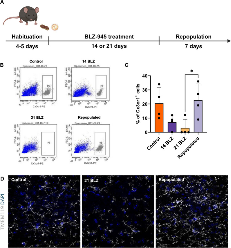
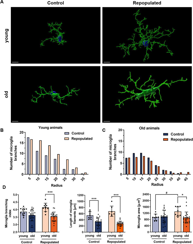
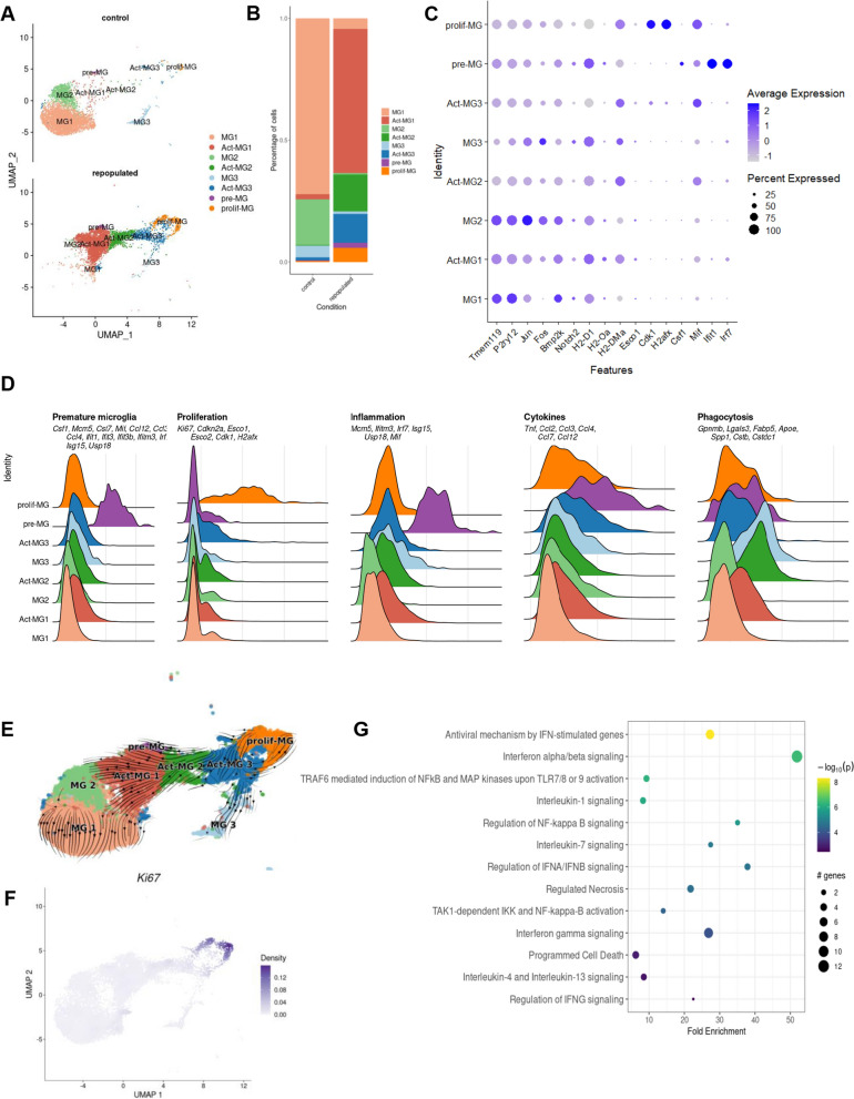
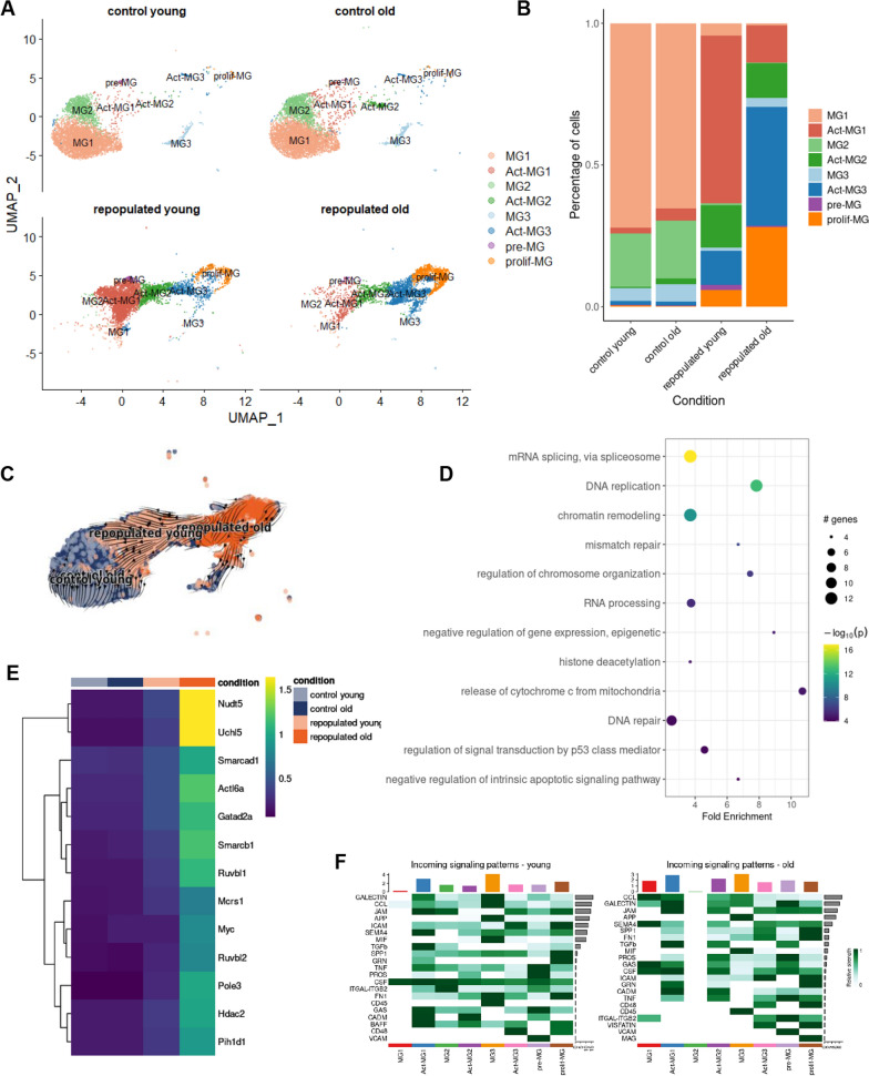
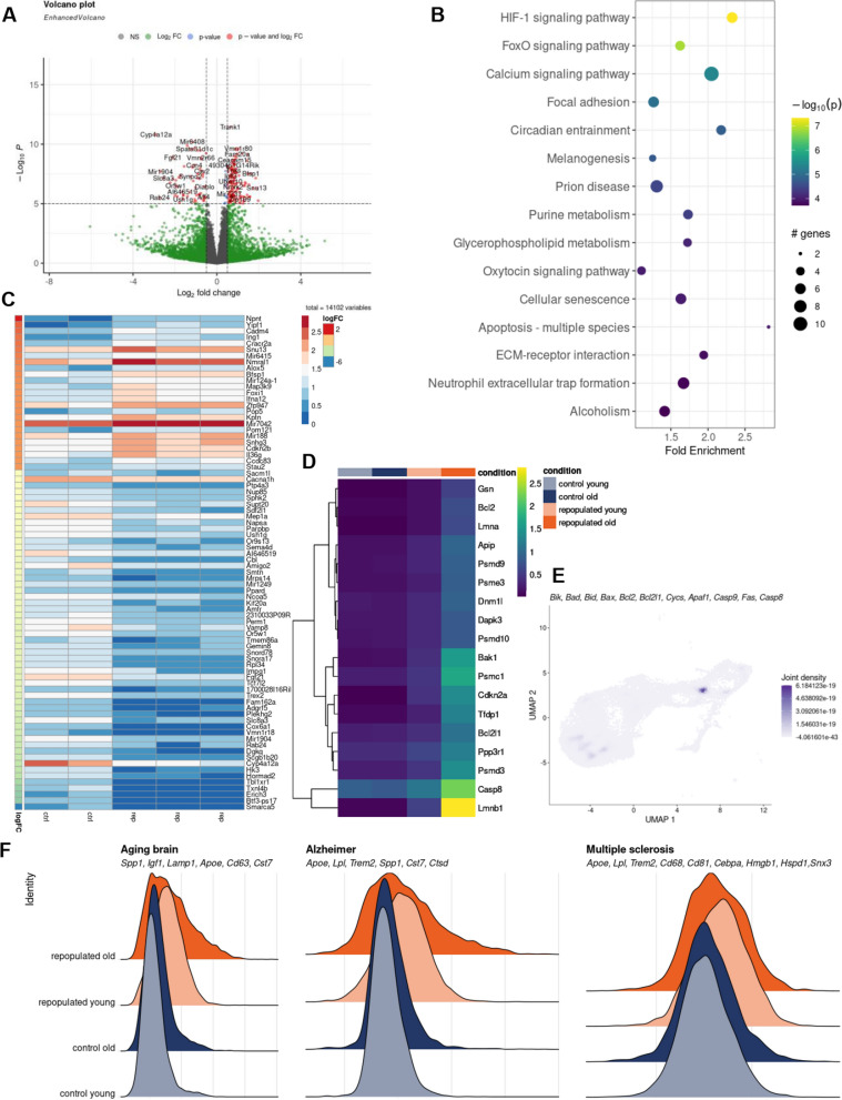
A CSRF1R inhibitor (BLZ-945) depleted microglia within 21 days and a number of microglia was fully restored within 7 days, as confirmed by TMEM119 staining and flow cytometry. ScRNA-seq and computational analyses demonstrate that repopulated microglia originated from preexisting progenitors and reconstituted functional clusters but upregulated inflammatory genes. Percentages of proliferating, immature microglia displaying inflammatory gene expression increased in aging mice. Morphometric analysis of MG cell body and branching revealed a distinct morphology of repopulated MG, particularly in brains of old mice. We demonstrate that with aging some repopulated MG fail to reach the homeostatic phenotype. These differences may contribute to the deterioration of MG protective functions with age.
小胶质细胞(MG)是中枢神经系统的髓系细胞,可支持内稳态,并在病变中引发神经炎症。单细胞 RNA 测序(scRNA-seq)揭示了小鼠大脑中 MG 的功能异质性。小胶质细胞是自我更新的细胞,集落刺激因子 1 受体(CSF1R)信号的抑制会消耗小胶质细胞,而小胶质细胞会迅速重新填充。重新填充的小胶质细胞的功能知之甚少。
我们结合了 scRNA-seq、批量 RNA-seq、免疫荧光和共聚焦成像,以研究重新填充的小胶质细胞的功能和形态。
CSF1R 抑制剂(BLZ-945)在 21 天内耗尽了小胶质细胞,而 TMEM119 染色和流式细胞术证实,在 7 天内,大量小胶质细胞得到了完全恢复。scRNA-seq 和计算分析表明,重新填充的小胶质细胞源自先前存在的祖细胞,并重新构建了功能性簇,但上调了炎症基因。在衰老小鼠中,增殖、不成熟的小胶质细胞中表达炎症基因的比例增加。对 MG 细胞体和分支的形态计量分析显示,重新填充的 MG 具有独特的形态,尤其是在老年小鼠的大脑中。我们证明,随着年龄的增长,一些重新填充的 MG 无法达到稳态表型。这些差异可能导致 MG 保护功能随年龄的恶化。