Institute of Cognitive Neuroscience, University College London, London, UK.
Department of Psychology & York Biomedical Research Institute, University of York, York, UK.
J Psychopharmacol. 2024 Dec;38(12):1071-1082. doi: 10.1177/02698811241286773. Epub 2024 Oct 4.
Selective serotonin reuptake inhibitors (SSRIs) are used for the treatment of several conditions including anxiety disorders, but the basic neurobiology of serotonin function remains unclear. The amygdala and prefrontal cortex are strongly innervated by serotonergic projections and have been suggested to play an important role in anxiety expression. However, serotonergic function in behaviour and SSRI-mediated neurobiological changes remain incompletely understood.
To investigate the neural correlates of subchronic antidepressant administration.
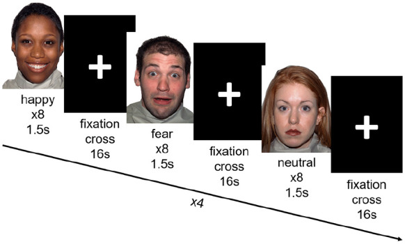
We investigated whether the 2- to 3-week administration of a highly selective SSRI (escitalopram) would alter brain activation on a task robustly shown to recruit the bilateral amygdala and frontal cortices in a large healthy volunteer sample. Participants performed the task during a functional magnetic resonance imaging acquisition before ( = 96) and after subchronic escitalopram ( = 46, days of administration mean (SD) = 15.7 (2.70)) or placebo ( = 40 days of administration mean (SD) = 16.2 (2.90)) self-administration.
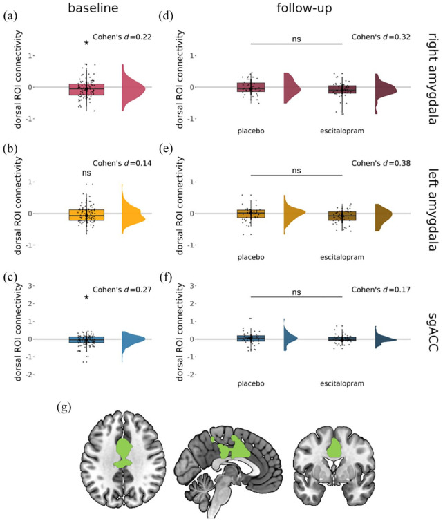
Compared to placebo, we found an elevation in right amygdala activation to the task after escitalopram administration without significant changes in mood. This effect was not seen in the left amygdala, the dorsomedial region of interest, the subgenual anterior cingulate cortex or the right fusiform area. There were no significant changes in connectivity between the dorsomedial cortex and amygdala or the subgenual anterior cingulate cortex after escitalopram administration.
To date, this most highly powered study of subchronic SSRI administration indicates that, contrary to effects often seen in patients with anxiety disorders, subchronic SSRI treatment may amygdala activation in healthy controls. This finding highlights important gaps in our understanding of the functional role of serotonin.
选择性 5-羟色胺再摄取抑制剂(SSRIs)被用于治疗多种疾病,包括焦虑症,但 5-羟色胺功能的基本神经生物学仍不清楚。杏仁核和前额叶皮层被强烈的 5-羟色胺能投射所支配,并被认为在焦虑表达中起重要作用。然而,5-羟色胺能功能在行为和 SSRI 介导的神经生物学变化方面仍不完全清楚。
研究亚慢性抗抑郁治疗的神经相关性。
我们调查了 2-3 周的高选择性 SSRI(依地普仑)给药是否会改变大脑在一项任务中的激活,该任务在一个大型健康志愿者样本中强有力地显示了双侧杏仁核和前额皮质的募集。参与者在接受功能磁共振成像采集之前(=96)和亚慢性依地普仑(=46,给药天数的平均值(SD)=15.7(2.70))或安慰剂(=40 天给药天数的平均值(SD)=16.2(2.90))自我给药后执行该任务。
与安慰剂相比,我们发现依地普仑给药后,右侧杏仁核对任务的激活增加,而情绪没有明显变化。这种效应在左侧杏仁核、内侧前额叶区域、扣带回下前部或右侧梭状回中没有看到。依地普仑给药后,内侧前额叶皮质与杏仁核或扣带回下前部之间的连接没有明显变化。
到目前为止,这项关于亚慢性 SSRI 给药的研究表明,与焦虑症患者经常出现的作用相反,亚慢性 SSRI 治疗可能会增加健康对照组的杏仁核激活。这一发现突出了我们对 5-羟色胺功能的理解中的重要差距。