• 文献检索
  • 文档翻译
  • 深度研究
  • 学术资讯
  • Suppr Zotero 插件Zotero 插件
  • 邀请有礼
  • 套餐&价格
  • 历史记录
应用&插件
Suppr Zotero 插件Zotero 插件浏览器插件Mac 客户端Windows 客户端微信小程序
定价
高级版会员购买积分包购买API积分包
服务
文献检索文档翻译深度研究API 文档MCP 服务
关于我们
关于 Suppr公司介绍联系我们用户协议隐私条款
关注我们

Suppr 超能文献

核心技术专利:CN118964589B侵权必究
粤ICP备2023148730 号-1Suppr @ 2026

文献检索

告别复杂PubMed语法,用中文像聊天一样搜索,搜遍4000万医学文献。AI智能推荐,让科研检索更轻松。

立即免费搜索

文件翻译

保留排版,准确专业,支持PDF/Word/PPT等文件格式,支持 12+语言互译。

免费翻译文档

深度研究

AI帮你快速写综述,25分钟生成高质量综述,智能提取关键信息,辅助科研写作。

立即免费体验

一种灵活的生成算法,用于在计算机中生成胎盘。

A flexible generative algorithm for growing in silico placentas.

机构信息

Department of Mechanical Engineering, University College London, London, United Kingdom.

Centre for the Developing Brain, School of Biomedical Engineering and Imaging Sciences, King's College London, London, United Kingdom.

出版信息

PLoS Comput Biol. 2024 Oct 7;20(10):e1012470. doi: 10.1371/journal.pcbi.1012470. eCollection 2024 Oct.

DOI:10.1371/journal.pcbi.1012470
PMID:39374295
原文链接:https://pmc.ncbi.nlm.nih.gov/articles/PMC11486434/
Abstract

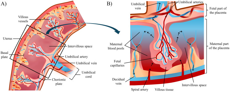
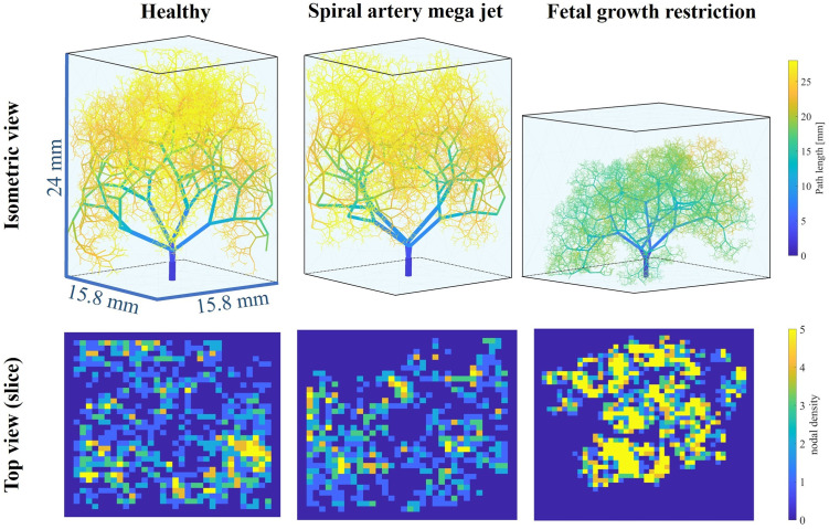
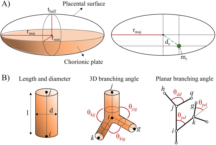
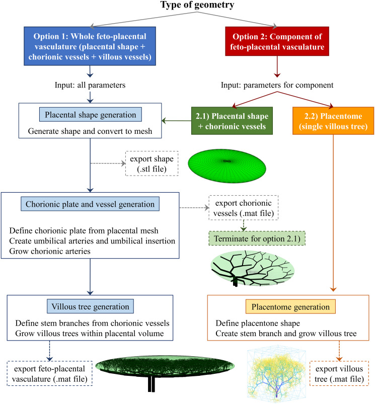
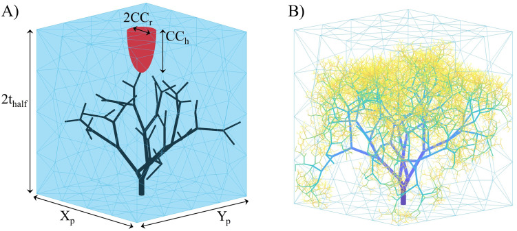
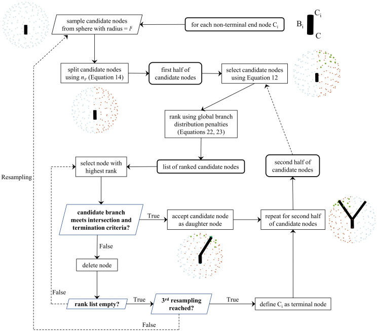
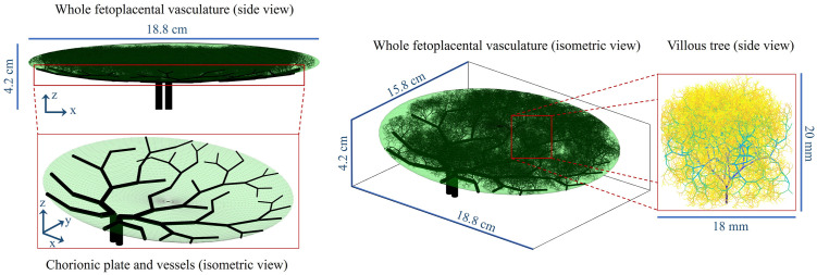
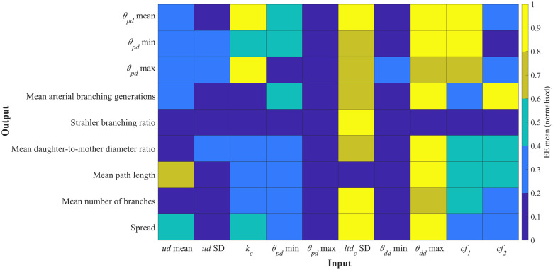
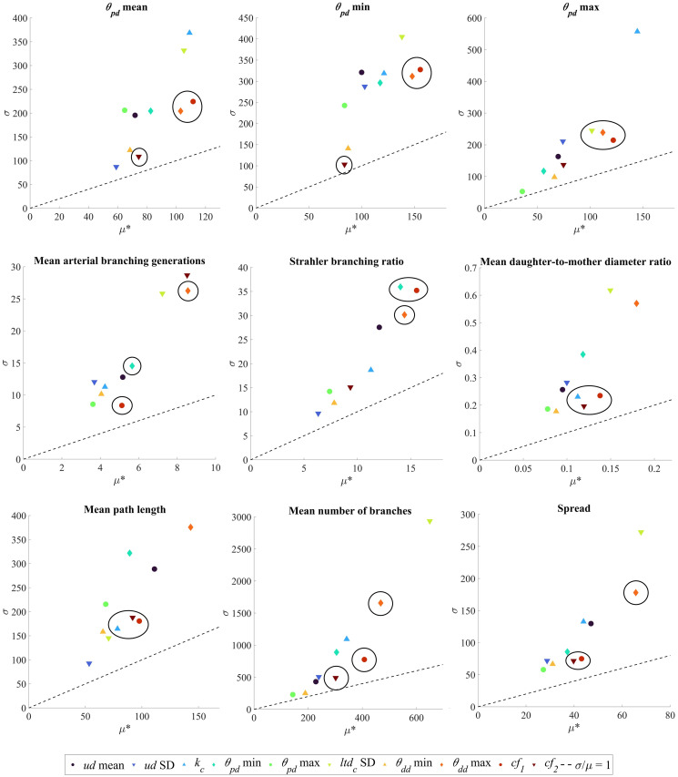
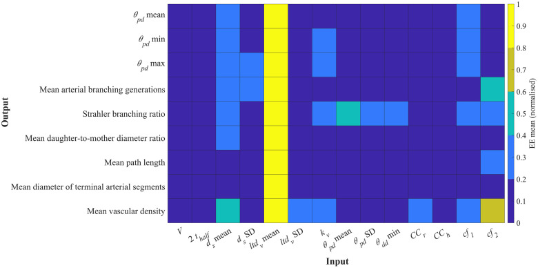
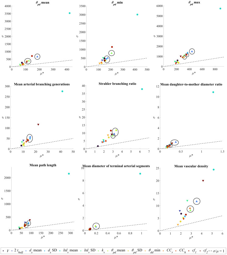
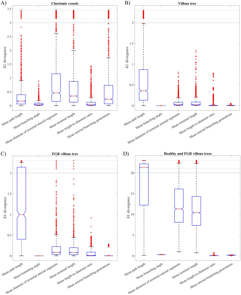
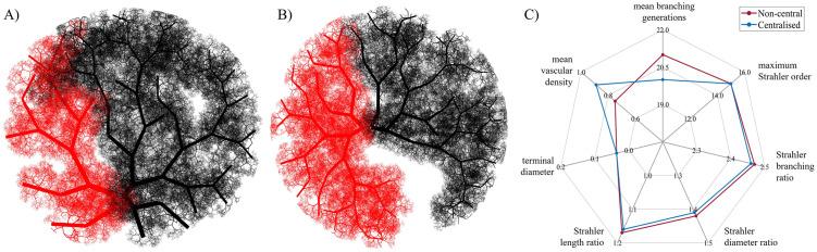
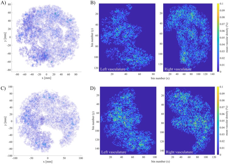
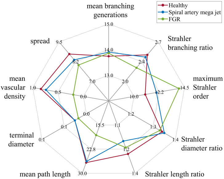
The placenta is crucial for a successful pregnancy, facilitating oxygen exchange and nutrient transport between mother and fetus. Complications like fetal growth restriction and pre-eclampsia are linked to placental vascular structure abnormalities, highlighting the need for early detection of placental health issues. Computational modelling offers insights into how vascular architecture correlates with flow and oxygenation in both healthy and dysfunctional placentas. These models use synthetic networks to represent the multiscale feto-placental vasculature, but current methods lack direct control over key morphological parameters like branching angles, essential for predicting placental dysfunction. We introduce a novel generative algorithm for creating in silico placentas, allowing user-controlled customisation of feto-placental vasculatures, both as individual components (placental shape, chorionic vessels, placentone) and as a complete structure. The algorithm is physiologically underpinned, following branching laws (i.e. Murray's Law), and is defined by four key morphometric statistics: vessel diameter, vessel length, branching angle and asymmetry. Our algorithm produces structures consistent with in vivo measurements and ex vivo observations. Our sensitivity analysis highlights how vessel length variations and branching angles play a pivotal role in defining the architecture of the placental vascular network. Moreover, our approach is stochastic in nature, yielding vascular structures with different topological metrics when imposing the same input settings. Unlike previous volume-filling algorithms, our approach allows direct control over key morphological parameters, generating vascular structures that closely resemble real vascular densities and allowing for the investigation of the impact of morphological parameters on placental function in upcoming studies.

摘要

胎盘对于成功妊娠至关重要,促进了母体和胎儿之间的氧气交换和营养物质运输。胎儿生长受限和子痫前期等并发症与胎盘血管结构异常有关,这凸显了早期发现胎盘健康问题的重要性。计算模型提供了关于血管结构如何与健康和功能失调胎盘中的血流和氧合相关联的深入了解。这些模型使用合成网络来表示多尺度的胎儿胎盘血管系统,但目前的方法缺乏对关键形态参数的直接控制,例如分支角度,这对于预测胎盘功能障碍至关重要。我们引入了一种新的生成算法,用于创建计算机模拟胎盘,允许用户控制胎儿胎盘血管系统的定制,包括单个组件(胎盘形状、绒毛膜血管、胎盘)和完整结构。该算法具有生理学基础,遵循分支定律(即默里定律),并由四个关键形态统计学定义:血管直径、血管长度、分支角度和不对称性。我们的算法生成的结构与体内测量和离体观察一致。我们的敏感性分析强调了血管长度变化和分支角度如何在定义胎盘血管网络结构中发挥关键作用。此外,我们的方法本质上是随机的,当施加相同的输入设置时,会产生具有不同拓扑度量的血管结构。与以前的体积填充算法不同,我们的方法可以直接控制关键形态参数,生成与真实血管密度非常相似的血管结构,并允许在即将进行的研究中调查形态参数对胎盘功能的影响。

https://cdn.ncbi.nlm.nih.gov/pmc/blobs/8d85/11486434/034104fe1365/pcbi.1012470.g015.jpg
https://cdn.ncbi.nlm.nih.gov/pmc/blobs/8d85/11486434/75c98ab34f64/pcbi.1012470.g001.jpg
https://cdn.ncbi.nlm.nih.gov/pmc/blobs/8d85/11486434/5a8d2e38a2dd/pcbi.1012470.g002.jpg
https://cdn.ncbi.nlm.nih.gov/pmc/blobs/8d85/11486434/9b65fd003023/pcbi.1012470.g003.jpg
https://cdn.ncbi.nlm.nih.gov/pmc/blobs/8d85/11486434/00cf8eb1c6be/pcbi.1012470.g004.jpg
https://cdn.ncbi.nlm.nih.gov/pmc/blobs/8d85/11486434/5a7312e3a0a0/pcbi.1012470.g005.jpg
https://cdn.ncbi.nlm.nih.gov/pmc/blobs/8d85/11486434/ee2fc77ad309/pcbi.1012470.g006.jpg
https://cdn.ncbi.nlm.nih.gov/pmc/blobs/8d85/11486434/e9552a4edde0/pcbi.1012470.g007.jpg
https://cdn.ncbi.nlm.nih.gov/pmc/blobs/8d85/11486434/0f8de68d8e2c/pcbi.1012470.g008.jpg
https://cdn.ncbi.nlm.nih.gov/pmc/blobs/8d85/11486434/16b4c519f6b7/pcbi.1012470.g009.jpg
https://cdn.ncbi.nlm.nih.gov/pmc/blobs/8d85/11486434/f86a910196f6/pcbi.1012470.g010.jpg
https://cdn.ncbi.nlm.nih.gov/pmc/blobs/8d85/11486434/d45e740c7751/pcbi.1012470.g011.jpg
https://cdn.ncbi.nlm.nih.gov/pmc/blobs/8d85/11486434/89ebef5cb199/pcbi.1012470.g012.jpg
https://cdn.ncbi.nlm.nih.gov/pmc/blobs/8d85/11486434/5aee0df17080/pcbi.1012470.g013.jpg
https://cdn.ncbi.nlm.nih.gov/pmc/blobs/8d85/11486434/f9817fcab5e2/pcbi.1012470.g014.jpg
https://cdn.ncbi.nlm.nih.gov/pmc/blobs/8d85/11486434/034104fe1365/pcbi.1012470.g015.jpg

相似文献

1
A flexible generative algorithm for growing in silico placentas.一种灵活的生成算法,用于在计算机中生成胎盘。
PLoS Comput Biol. 2024 Oct 7;20(10):e1012470. doi: 10.1371/journal.pcbi.1012470. eCollection 2024 Oct.
2
Structure-function relationships in the feto-placental circulation from in silico interpretation of micro-CT vascular structures.基于微计算机断层扫描血管结构的计算机模拟解释,探讨胎儿-胎盘循环中的结构-功能关系。
J Theor Biol. 2021 May 21;517:110630. doi: 10.1016/j.jtbi.2021.110630. Epub 2021 Feb 17.
3
Quantitative characterization of rodent feto-placental vasculature morphology in micro-computed tomography images.定量分析微计算机断层扫描图像中小鼠胎仔胎盘血管形态。
Comput Methods Programs Biomed. 2019 Oct;179:104984. doi: 10.1016/j.cmpb.2019.104984. Epub 2019 Jul 15.
4
Three-dimensional flow patterns in the feto-placental vasculature system of the mouse placenta.小鼠胎盘胎儿-胎盘血管系统中的三维血流模式。
Microvasc Res. 2017 May;111:88-95. doi: 10.1016/j.mvr.2017.01.004. Epub 2017 Jan 19.
5
Investigating the Upstream and Downstream Hemodynamic Boundary Conditions of Healthy and Growth-Restricted Rat Feto-Placental Arterial Networks.研究健康和生长受限的大鼠胎-胎盘动脉网络的上下游血流动力学边界条件。
Ann Biomed Eng. 2021 Sep;49(9):2183-2195. doi: 10.1007/s10439-021-02749-4. Epub 2021 Mar 1.
6
Quantitative 3D micro-CT imaging of the human feto-placental vasculature in intrauterine growth restriction.宫内生长受限中人类胎儿-胎盘血管系统的定量三维显微CT成像
Placenta. 2008 Nov;29(11):937-41. doi: 10.1016/j.placenta.2008.08.017. Epub 2008 Oct 11.
7
Assessment of first-trimester utero-placental vascular morphology by 3D power Doppler ultrasound image analysis using a skeletonization algorithm: the Rotterdam Periconception Cohort.基于骨架化算法的三维能量多普勒超声图像分析对早孕期子宫胎盘血管形态的评估:鹿特丹围孕期队列研究。
Hum Reprod. 2022 Oct 31;37(11):2532-2545. doi: 10.1093/humrep/deac202.
8
Morphologic development of the first-trimester utero-placental vasculature is positively associated with embryonic and fetal growth: the Rotterdam Periconception Cohort.头三个月的子宫胎盘血管形态发育与胚胎和胎儿生长呈正相关:鹿特丹围孕期队列研究。
Hum Reprod. 2024 May 2;39(5):923-935. doi: 10.1093/humrep/deae056.
9
Multiscale modelling of the feto-placental vasculature.胎儿-胎盘血管系统的多尺度建模。
Interface Focus. 2015 Apr 6;5(2):20140078. doi: 10.1098/rsfs.2014.0078.
10
A review of feto-placental vasculature flow modelling.胎儿-胎盘血管血流建模综述。
Placenta. 2023 Oct;142:56-63. doi: 10.1016/j.placenta.2023.08.068. Epub 2023 Aug 22.

本文引用的文献

1
Reconstructing microvascular network skeletons from 3D images: What is the ground truth?从 3D 图像重建微血管网络骨架:什么是真实情况?
Comput Biol Med. 2024 Mar;171:108140. doi: 10.1016/j.compbiomed.2024.108140. Epub 2024 Feb 27.
2
Blood flow and transport in the human placenta.人体胎盘的血流与物质运输
Annu Rev Fluid Mech. 2019 Jan;51:25-47. doi: 10.1146/annurev-fluid-010518-040219. Epub 2018 Aug 3.
3
Pregnancy Complications, Correlation With Placental Pathology and Neonatal Outcomes.妊娠并发症、与胎盘病理学及新生儿结局的相关性
Front Clin Diabetes Healthc. 2022 Mar 8;2:807192. doi: 10.3389/fcdhc.2021.807192. eCollection 2021.
4
Impaired placental hemodynamics and function in a non-human primate model of gestational protein restriction.妊娠蛋白限制的非人类灵长类动物模型中的胎盘血液动力学和功能障碍。
Sci Rep. 2023 Jan 16;13(1):841. doi: 10.1038/s41598-023-28051-y.
5
Quantitative longitudinal T2* mapping for assessing placental function and association with adverse pregnancy outcomes across gestation.定量纵向 T2* 映射评估胎盘功能与妊娠不良结局的相关性。
PLoS One. 2022 Jul 19;17(7):e0270360. doi: 10.1371/journal.pone.0270360. eCollection 2022.
6
Challenges and opportunities of integrating imaging and mathematical modelling to interrogate biological processes.整合成像和数学建模以探究生物过程的挑战和机遇。
Int J Biochem Cell Biol. 2022 May;146:106195. doi: 10.1016/j.biocel.2022.106195. Epub 2022 Mar 25.
7
A massively multi-scale approach to characterizing tissue architecture by synchrotron micro-CT applied to the human placenta.基于同步辐射微计算机断层扫描的大规模多尺度方法对人胎盘组织架构进行特征描述。
J R Soc Interface. 2021 Jun;18(179):20210140. doi: 10.1098/rsif.2021.0140. Epub 2021 Jun 2.
8
Adaptive constrained constructive optimisation for complex vascularisation processes.自适应约束构造优化在复杂血管化过程中的应用。
Sci Rep. 2021 Mar 17;11(1):6180. doi: 10.1038/s41598-021-85434-9.
9
Investigating the Upstream and Downstream Hemodynamic Boundary Conditions of Healthy and Growth-Restricted Rat Feto-Placental Arterial Networks.研究健康和生长受限的大鼠胎-胎盘动脉网络的上下游血流动力学边界条件。
Ann Biomed Eng. 2021 Sep;49(9):2183-2195. doi: 10.1007/s10439-021-02749-4. Epub 2021 Mar 1.
10
Structure-function relationships in the feto-placental circulation from in silico interpretation of micro-CT vascular structures.基于微计算机断层扫描血管结构的计算机模拟解释,探讨胎儿-胎盘循环中的结构-功能关系。
J Theor Biol. 2021 May 21;517:110630. doi: 10.1016/j.jtbi.2021.110630. Epub 2021 Feb 17.