Savoldi Anna Paola, Anghileri Elena, Moscatelli Marco, Silvani Antonio, Pollo Bianca, Valeria Cuccarini, Pascuzzo Riccardo, Aquino Domenico, Grisoli Marina, Doniselli Fabio M
Neuroradiology Unit, Fondazione IRCCS Istituto Neurologico Carlo Besta, I-20133 Milan, Italy.
Neuro-Oncology Unit, Fondazione IRCCS Istituto Neurologico Carlo Besta, I-20133 Milan, Italy.
Oncol Lett. 2024 Sep 27;28(6):570. doi: 10.3892/ol.2024.14703. eCollection 2024 Dec.
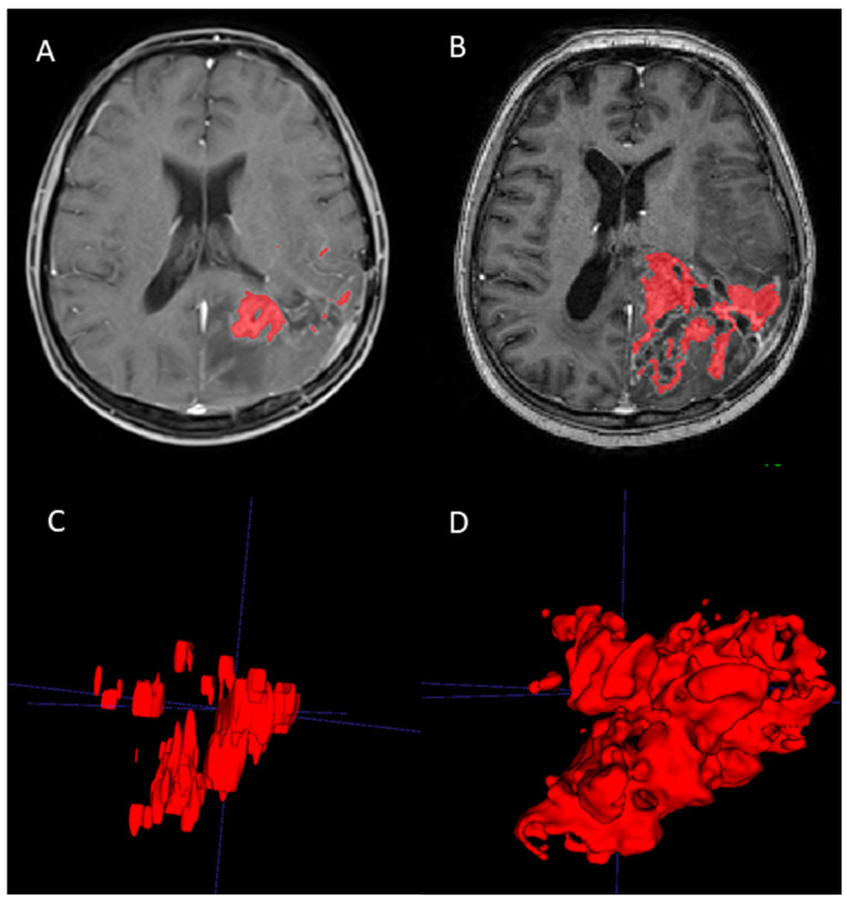
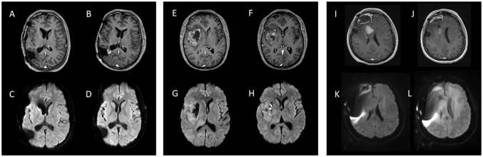
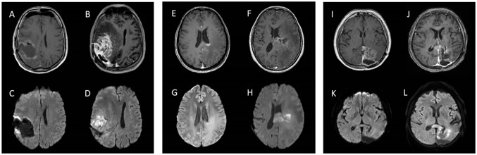
The use of fotemustine (FTM) has been authorized in certain countries for the treatment of recurrent high-grade gliomas (HGG) after Stupp therapy. However, to the best of our knowledge, no studies have assessed changes in magnetic resonance imaging (MRI) during treatment with FTM monotherapy. The aim of the present study was to assess the neuroradiological findings in a cohort of patients with recurrent HGG treated with FTM monotherapy. Patients with HGG already undergoing the Stupp protocol were retrospectively included. MRIs (pre- and post-FTM treatment) were analyzed by two neuroradiologists in consensus: Volume and diffusion values of the contrast-enhanced component were measured on T1-weighted volumetric sequences after gadolinium injection and on apparent diffusion coefficient (ADC) maps, respectively. A total of 19 patients [median age, 49 years; interquartile range (IQR), 43-57 years] were included, 17 of whom had glioblastoma and 2 had astrocytoma isocitrate dehydrogenase-mutated grade 4. The median duration of FTM therapy was 4 months (IQR, 2-6 months). The median tumor volume measured on the contrast-enhanced component was 2,216 mm (IQR, 768-13,169 mm) at baseline and 9,217 mm (IQR, 3,455-16,697 mm) at the end of treatment, with a median change of +38% (IQR, -45-+574%). A total of seven patients showed a volume decrease. ADC value analysis of the enhancement area demonstrated no significant difference between the pre- and the post-FTM treatment periods (P=0.36); however, in three patients, the decreases in ADC levels were particularly marked. In conclusion, the present study described a series of patients with recurrent HGG treated with FTM in monotherapy, demonstrating a prevalent increase in lesion enhancement and three cases of marked restrictions on diffusion-weighted imaging. Further prospective studies are required to corroborate such preliminary results.
福莫司汀(FTM)在某些国家已被批准用于治疗经Stupp方案治疗后的复发性高级别胶质瘤(HGG)。然而,据我们所知,尚无研究评估FTM单药治疗期间磁共振成像(MRI)的变化。本研究的目的是评估接受FTM单药治疗的复发性HGG患者队列的神经放射学表现。回顾性纳入已接受Stupp方案治疗的HGG患者。两名神经放射科医生共同分析MRI(FTM治疗前后):分别在注射钆后T1加权容积序列和表观扩散系数(ADC)图上测量强化成分的体积和扩散值。共纳入19例患者[中位年龄49岁;四分位间距(IQR),43 - 57岁],其中17例为胶质母细胞瘤,2例为异柠檬酸脱氢酶突变的4级星形细胞瘤。FTM治疗的中位持续时间为4个月(IQR,2 - 6个月)。强化成分测量的肿瘤体积中位数在基线时为2216 mm³(IQR,768 - 13169 mm³),治疗结束时为9217 mm³(IQR,3455 - 16697 mm³),中位变化为 +38%(IQR, -45 - +574%)。共有7例患者体积减小。强化区域的ADC值分析显示FTM治疗前后无显著差异(P = 0.36);然而,3例患者的ADC水平下降尤为明显。总之,本研究描述了一组接受FTM单药治疗的复发性HGG患者,显示病变强化普遍增加,3例在扩散加权成像上有明显受限。需要进一步的前瞻性研究来证实这些初步结果。