• 文献检索
  • 文档翻译
  • 深度研究
  • 学术资讯
  • Suppr Zotero 插件Zotero 插件
  • 邀请有礼
  • 套餐&价格
  • 历史记录
应用&插件
Suppr Zotero 插件Zotero 插件浏览器插件Mac 客户端Windows 客户端微信小程序
定价
高级版会员购买积分包购买API积分包
服务
文献检索文档翻译深度研究API 文档MCP 服务
关于我们
关于 Suppr公司介绍联系我们用户协议隐私条款
关注我们

Suppr 超能文献

核心技术专利:CN118964589B侵权必究
粤ICP备2023148730 号-1Suppr @ 2026

文献检索

告别复杂PubMed语法,用中文像聊天一样搜索,搜遍4000万医学文献。AI智能推荐,让科研检索更轻松。

立即免费搜索

文件翻译

保留排版,准确专业,支持PDF/Word/PPT等文件格式,支持 12+语言互译。

免费翻译文档

深度研究

AI帮你快速写综述,25分钟生成高质量综述,智能提取关键信息,辅助科研写作。

立即免费体验

多组学分析确定肝硬化肝细胞癌的分子机制。

Multi-omic analysis identifies the molecular mechanism of hepatocellular carcinoma with cirrhosis.

机构信息

Department of Infectious Diseases, The First Affiliated Hospital of Zhengzhou University, Zhengzhou, 450052, Henan, China.

Department of Oncology, The First Affiliated Hospital, College of Clinical Medicine, Henan University of Science and Technology, Luoyang, 471000, Henan, China.

出版信息

Sci Rep. 2024 Oct 11;14(1):23832. doi: 10.1038/s41598-024-75609-5.

DOI:10.1038/s41598-024-75609-5
PMID:39394373
原文链接:https://pmc.ncbi.nlm.nih.gov/articles/PMC11470084/
Abstract

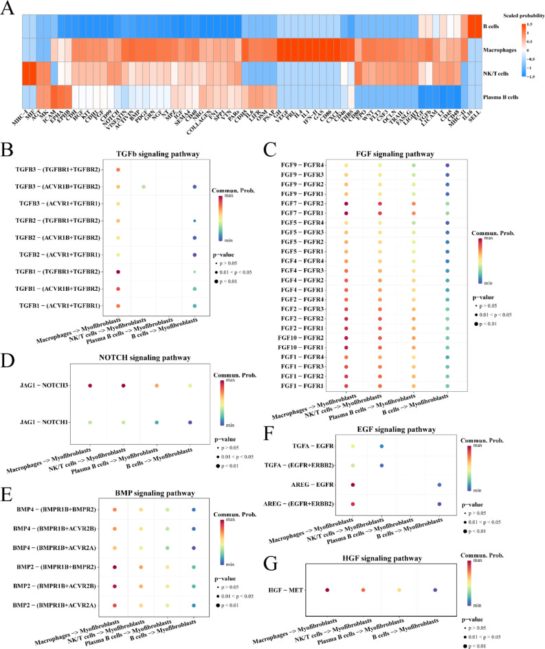
Hepatocellular carcinoma with cirrhosis promotes the advancement of malignancy and the development of fibrosis in normal liver tissues. Understanding the pathological mechanisms underlying the development of HCC with cirrhosis is important for developing effective therapeutic strategies. Herein, the RNA-sequencing (RNA-seq) data and corresponding clinical features of patients with HCC were extracted from The Cancer Genome Atlas (TCGA) database using the University of California Santa Cruz (UCSC) Xena platform. The enrichment degree of hallmarkers for each TCGA-LIHC cohort was quantified by ssGSEA algorithm. Weighted gene co-expression network analysis (WGCNA) revealed two gene module eigengenes (MEs) associated with cirrhosis, namely, MEbrown and MEgreen. Analysis of these modules using AUCell showed that MEbrown had higher enrichment scores in all immune cells, whereas MEgreen had higher enrichment scores in malignant cells. The CellChat package revealed that both immune and malignant cells contributed to the fibrotic activity of myofibroblasts through diverse signaling pathways. Additionally, spatial transcriptomic data showed that hepatocytes, proliferating hepatocytes, macrophages, and myofibroblasts were located in closer proximity in HCC tissues. These cells may potentially participate in the process of stimulating myofibroblast fibrotic activity, which may be related to the development of liver fibrosis. In summary, we made full use of multi-omics data to explore gene networks and cell types that may be involved in the development and progression of cirrhosis in HCC.

摘要

伴有肝硬化的肝细胞癌促进了恶性肿瘤的进展和正常肝组织的纤维化发展。了解伴有肝硬化的 HCC 发展的病理机制对于开发有效的治疗策略非常重要。在此,使用加利福尼亚大学圣克鲁兹分校(UCSC)Xena 平台从癌症基因组图谱(TCGA)数据库中提取了 HCC 患者的 RNA 测序(RNA-seq)数据和相应的临床特征。通过 ssGSEA 算法量化了每个 TCGA-LIHC 队列 hallmarker 的富集程度。加权基因共表达网络分析(WGCNA)揭示了与肝硬化相关的两个基因模块特征基因(ME),即 MEbrown 和 MEgreen。使用 AUCell 对这些模块进行分析表明,MEbrown 在所有免疫细胞中的富集评分更高,而 MEgreen 在恶性细胞中的富集评分更高。CellChat 包表明,免疫细胞和恶性细胞均通过多种信号通路促进成纤维细胞肌成纤维细胞的纤维化活性。此外,空间转录组学数据表明,肝癌组织中的肝细胞、增殖肝细胞、巨噬细胞和成纤维细胞彼此更接近。这些细胞可能潜在地参与刺激成纤维细胞肌成纤维细胞的纤维化活性的过程,这可能与肝纤维化的发展有关。总之,我们充分利用多组学数据来探索可能参与 HCC 中肝硬化发展和进展的基因网络和细胞类型。

https://cdn.ncbi.nlm.nih.gov/pmc/blobs/fd6f/11470084/1280a3b1046f/41598_2024_75609_Fig7_HTML.jpg
https://cdn.ncbi.nlm.nih.gov/pmc/blobs/fd6f/11470084/fc4bffdd9ac8/41598_2024_75609_Fig1_HTML.jpg
https://cdn.ncbi.nlm.nih.gov/pmc/blobs/fd6f/11470084/1fe8a818655c/41598_2024_75609_Fig2_HTML.jpg
https://cdn.ncbi.nlm.nih.gov/pmc/blobs/fd6f/11470084/7b32965586e7/41598_2024_75609_Fig3_HTML.jpg
https://cdn.ncbi.nlm.nih.gov/pmc/blobs/fd6f/11470084/0c9793605de3/41598_2024_75609_Fig4_HTML.jpg
https://cdn.ncbi.nlm.nih.gov/pmc/blobs/fd6f/11470084/1bf8ba91651a/41598_2024_75609_Fig5_HTML.jpg
https://cdn.ncbi.nlm.nih.gov/pmc/blobs/fd6f/11470084/36b2d8dcd7b1/41598_2024_75609_Fig6_HTML.jpg
https://cdn.ncbi.nlm.nih.gov/pmc/blobs/fd6f/11470084/1280a3b1046f/41598_2024_75609_Fig7_HTML.jpg

相似文献

1
Multi-omic analysis identifies the molecular mechanism of hepatocellular carcinoma with cirrhosis.多组学分析确定肝硬化肝细胞癌的分子机制。
Sci Rep. 2024 Oct 11;14(1):23832. doi: 10.1038/s41598-024-75609-5.
2
Developing an advanced diagnostic model for hepatocellular carcinoma through multi-omics integration leveraging diverse cell-death patterns.通过整合多种细胞死亡模式的多组学方法开发用于肝细胞癌的先进诊断模型。
Front Immunol. 2024 Jul 9;15:1410603. doi: 10.3389/fimmu.2024.1410603. eCollection 2024.
3
Identification of cancer stem cell characteristics in liver hepatocellular carcinoma by WGCNA analysis of transcriptome stemness index.通过转录组干性指数的 WGCNA 分析鉴定肝癌中的癌症干细胞特征。
Cancer Med. 2020 Jun;9(12):4290-4298. doi: 10.1002/cam4.3047. Epub 2020 Apr 20.
4
Paracrine effects of CCN3 from non-cancerous hepatic cells increase signaling and progression of hepatocellular carcinoma.非癌性肝细胞分泌的 CCN3 通过旁分泌作用增加信号传导并促进肝癌进展。
BMC Cancer. 2019 Apr 27;19(1):395. doi: 10.1186/s12885-019-5603-7.
5
Prognostic value of long non-coding RNA in hepatocellular carcinoma: A study based on multi-omics analysis and RT-PCR validation.长链非编码 RNA 在肝细胞癌中的预后价值:基于多组学分析和 RT-PCR 验证的研究。
Pathol Oncol Res. 2023 Jan 4;28:1610808. doi: 10.3389/pore.2022.1610808. eCollection 2022.
6
Functional and topological properties in hepatocellular carcinoma transcriptome.肝细胞癌转录组中的功能和拓扑性质。
PLoS One. 2012;7(4):e35510. doi: 10.1371/journal.pone.0035510. Epub 2012 Apr 23.
7
Bioinformatics analysis of key genes and pathways for hepatocellular carcinoma transformed from cirrhosis.肝硬化转化为肝细胞癌关键基因和通路的生物信息学分析
Medicine (Baltimore). 2017 Jun;96(25):e6938. doi: 10.1097/MD.0000000000006938.
8
An Immune Gene Expression Signature Associated With Development of Human Hepatocellular Carcinoma Identifies Mice That Respond to Chemopreventive Agents.与人类肝细胞癌发展相关的免疫基因表达谱可识别对化学预防剂有反应的小鼠。
Gastroenterology. 2019 Nov;157(5):1383-1397.e11. doi: 10.1053/j.gastro.2019.07.028. Epub 2019 Jul 22.
9
Multi-omic approaches for characterization of hepatocellular carcinoma.用于肝细胞癌特征描述的多组学方法
Annu Int Conf IEEE Eng Med Biol Soc. 2016 Aug;2016:3437-3440. doi: 10.1109/EMBC.2016.7591467.
10
Integrative analysis unveils ECM signatures and pathways driving hepatocellular carcinoma progression: A multi-omics approach and prognostic model development.整合分析揭示了 ECM 特征和驱动肝细胞癌进展的途径:一种多组学方法和预后模型的开发。
J Cell Mol Med. 2024 Apr;28(8):e18230. doi: 10.1111/jcmm.18230.

本文引用的文献

1
The hepatocyte epidermal growth factor receptor (EGFR) pathway regulates the cellular interactome within the liver fibrotic niche.肝细胞表皮生长因子受体 (EGFR) 通路调节肝纤维化微环境中的细胞相互作用组。
J Pathol. 2024 Aug;263(4-5):482-495. doi: 10.1002/path.6299. Epub 2024 Jun 14.
2
Dibutyl phthalate (DBP) promotes Epithelial-Mesenchymal Transition (EMT) to aggravate liver fibrosis into cirrhosis and portal hypertension (PHT) via ROS/TGF-β1/Snail-1 signalling pathway in adult rats.邻苯二甲酸二丁酯(DBP)通过 ROS/TGF-β1/Snail-1 信号通路促进成年大鼠的上皮-间充质转化(EMT),从而加重肝纤维化向肝硬化和门静脉高压(PHT)的发展。
Ecotoxicol Environ Saf. 2024 Apr 1;274:116124. doi: 10.1016/j.ecoenv.2024.116124. Epub 2024 Mar 18.
3
Distinct fibroblast subpopulations associated with bone, brain or intrapulmonary metastasis in advanced non-small-cell lung cancer.
晚期非小细胞肺癌中与骨、脑或肺内转移相关的不同成纤维细胞亚群。
Clin Transl Med. 2024 Mar;14(3):e1605. doi: 10.1002/ctm2.1605.
4
Big data analytics for MerTK genomics reveals its double-edged sword functions in human diseases.大数据分析 MerTK 基因组学揭示其在人类疾病中的双刃剑功能。
Redox Biol. 2024 Apr;70:103061. doi: 10.1016/j.redox.2024.103061. Epub 2024 Feb 5.
5
EGFR-activated myofibroblasts promote metastasis of pancreatic cancer.表皮生长因子受体激活的肌成纤维细胞促进胰腺癌转移。
Cancer Cell. 2024 Jan 8;42(1):101-118.e11. doi: 10.1016/j.ccell.2023.12.002. Epub 2023 Dec 28.
6
Exploration and validation of key genes associated with early lymph node metastasis in thyroid carcinoma using weighted gene co-expression network analysis and machine learning.采用加权基因共表达网络分析和机器学习技术探索和验证甲状腺癌早期淋巴结转移相关的关键基因。
Front Endocrinol (Lausanne). 2023 Dec 8;14:1247709. doi: 10.3389/fendo.2023.1247709. eCollection 2023.
7
Use of albumin infusion for cirrhosis-related complications: An international position statement.白蛋白输注在肝硬化相关并发症中的应用:一项国际立场声明。
JHEP Rep. 2023 May 5;5(8):100785. doi: 10.1016/j.jhepr.2023.100785. eCollection 2023 Aug.
8
Identification of a novel signature based on macrophage-related marker genes to predict prognosis and immunotherapeutic effects in hepatocellular carcinoma.基于巨噬细胞相关标记基因鉴定一种新的特征以预测肝细胞癌的预后和免疫治疗效果。
Front Oncol. 2023 May 25;13:1176572. doi: 10.3389/fonc.2023.1176572. eCollection 2023.
9
Exosomes in liver fibrosis: The role of modulating hepatic stellate cells and immune cells, and prospects for clinical applications.外泌体在肝纤维化中的作用:调节肝星状细胞和免疫细胞的作用及临床应用前景。
Front Immunol. 2023 Mar 20;14:1133297. doi: 10.3389/fimmu.2023.1133297. eCollection 2023.
10
The evolving tumor microenvironment: From cancer initiation to metastatic outgrowth.不断演变的肿瘤微环境:从癌症起始到转移灶生长
Cancer Cell. 2023 Mar 13;41(3):374-403. doi: 10.1016/j.ccell.2023.02.016.