Tauziède-Espariat Arnault, Ebrahimi Azadeh, Boddaert Nathalie, Pietsch Torsten, Grajkowska Wieslawa, Blau Tobias, Koch Arend, Sievers Philipp, Guillemot Delphine, Pierron Gaëlle, Uro-Coste Emmanuelle, Nicaise Yvan, Siegfried Aurore, Gilles Adam, Bielle Franck, Mokhtari Karima, Cazals-Hatem Dominique, Iakovlev Gueorgui, Lhermitte Benoît, Entz-Werle Natacha, Csanyi Marie, Maurage Claude-Alain, Legrand Victor, Boutonnat Jean, Godfraind Catherine, McLeer Anne, Hasty Lauren, Métais Alice, Aboubakr Oumaima, Blauwblomme Thomas, Beccaria Kévin, Dangouloff-Ros Volodia, Varlet Pascale
Department of Neuropathology, GHU Paris-Psychiatrie et Neurosciences, Sainte-Anne Hospital, Paris, France.
INSERM U1266, IMABrain, Institute of Psychiatry and Neuroscience of Paris (IPNP), Université Paris Cité, Paris, France.
Brain Pathol. 2025 Mar;35(2):e13303. doi: 10.1111/bpa.13303. Epub 2024 Oct 23.
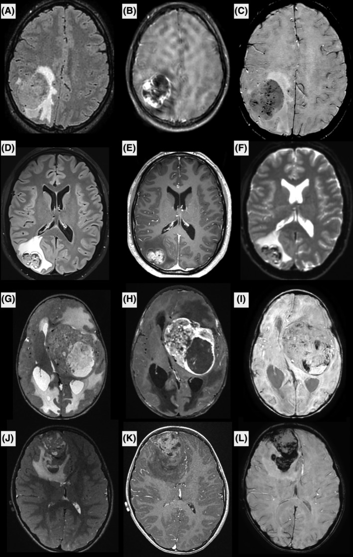
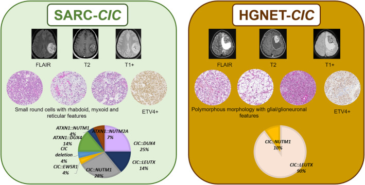
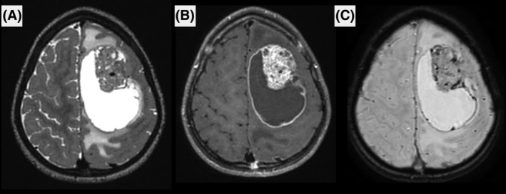
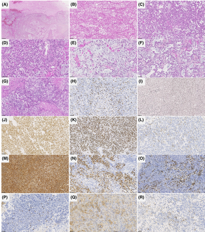
CIC fusions have been described in two different central nervous system (CNS) tumor entities. On one hand, fusions of CIC or ATXN1 genes belonging to the same complex of transcriptional repressors, were reported in the CIC-rearranged, sarcoma (SARC-CIC). The diagnosis of this tumor type, which was recently added to the World Health Organization (WHO) Classification of CNS tumors, is difficult mainly because the data concerning its histopathology (as compared to its soft tissue counterpart), immunoprofile, and clinical as well as radiological characteristics are scarce in the literature. On the other hand, a recent study, based on DNA-methylation profiling, has identified a novel high-grade neuroepithelial tumor characterized by recurrent CIC fusions (HGNET-CIC). The aim of this multicentric study was to characterize a cohort of 15 primary CNS tumors harboring a CIC or ATXN1 fusion in terms of clinical, radiological, histopathological, immunophenotypical, and epigenetic characteristics. According to the integrated diagnoses, 14/15 tumors corresponded to SARC-CIC, and only one to HGNET-CIC. The tumors showed similar clinical (mainly pediatric), radiological (mostly supratentorial, cystic, and contrast enhancing), immunophenotypical (common expression of glioneuronal markers), and genetic (similar spectrum of fusions) profiles but their histopathological appearance was clearly distinct. Moreover, we found a novel fusion transcript (CIC::EWSR1) in a SARC-CIC. Most DNA methylation profiles using the Heidelberg Brain Tumor Classifier (v12.8) annotated the samples to the methylation class "SARC-CIC" (9/14 tumors with available data). By using uniform manifold approximation and projection analysis, four other samples were classified as SARC-CIC and another clustered within the methylation class of HGNET-CIC. Our findings confirm that CNS CIC-fused tumors do not represent a single molecular tumor entity. Further analyses are needed to characterize HGNET-CIC in more detail. These results may help to refine the essential diagnostic criteria for SARC-CIC and their terminology (with a suggested consensual name of sarcoma, CIC/ATXN1-complex rearranged).
CIC融合已在两种不同的中枢神经系统(CNS)肿瘤实体中被描述。一方面,在CIC重排肉瘤(SARC-CIC)中报道了属于同一转录抑制复合物的CIC或ATXN1基因的融合。这种肿瘤类型最近被纳入世界卫生组织(WHO)中枢神经系统肿瘤分类,其诊断困难,主要是因为关于其组织病理学(与软组织对应物相比)、免疫表型以及临床和放射学特征的文献资料稀缺。另一方面,最近一项基于DNA甲基化谱分析的研究,确定了一种以复发性CIC融合为特征的新型高级别神经上皮肿瘤(HGNET-CIC)。这项多中心研究的目的是从临床、放射学、组织病理学、免疫表型和表观遗传学特征方面,对15例携带CIC或ATXN1融合的原发性中枢神经系统肿瘤进行特征描述。根据综合诊断,15例肿瘤中有14例符合SARC-CIC,只有1例符合HGNET-CIC。这些肿瘤显示出相似的临床(主要为儿童)、放射学(大多位于幕上、囊性且有强化)、免疫表型(神经胶质神经元标志物的共同表达)和遗传学(相似的融合谱)特征,但它们的组织病理学表现明显不同。此外,我们在一例SARC-CIC中发现了一种新的融合转录本(CIC::EWSR1)。使用海德堡脑肿瘤分类器(v12.8)的大多数DNA甲基化谱将样本注释为甲基化类别“SARC-CIC”(14例有可用数据的肿瘤中有9例)。通过使用均匀流形近似和投影分析,另外4个样本被分类为SARC-CIC,另一个聚类在HGNET-CIC的甲基化类别中。我们的研究结果证实,中枢神经系统CIC融合肿瘤并不代表单一的分子肿瘤实体。需要进一步分析以更详细地描述HGNET-CIC。这些结果可能有助于完善SARC-CIC的基本诊断标准及其术语(建议统一命名为肉瘤,CIC/ATXN1复合体重排)。