Schulz-Hildebrandt Hinnerk, Spasic Svetolik, Hou Fang, Ting Kuan-Chung, Batts Shelley, Tearney Guillermo, Stankovic Konstantina M
Wellman Center for Photomedicine, Massachusetts General Hospital, Harvard Medical School, Boston, MA, United States.
Department of Otolaryngology-Head and Neck Surgery, Stanford University School of Medicine, Stanford, CA, United States.
Front Mol Neurosci. 2024 Oct 10;17:1436837. doi: 10.3389/fnmol.2024.1436837. eCollection 2024.
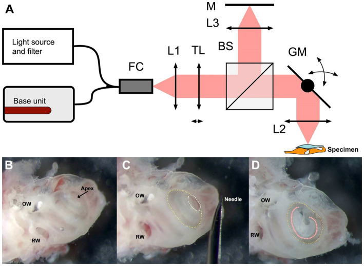
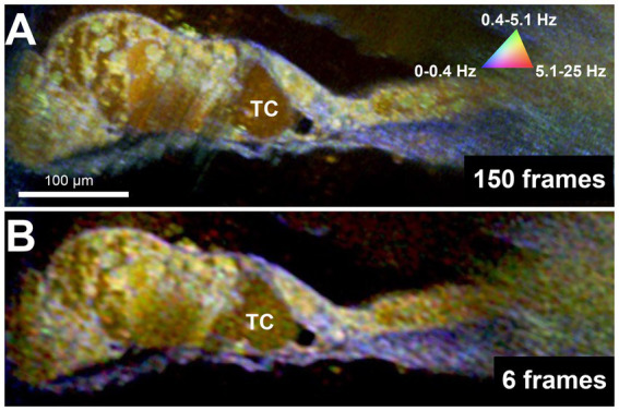
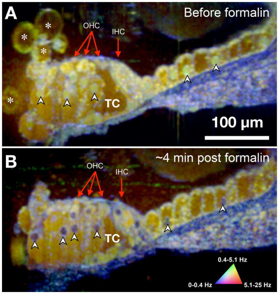
Sensorineural hearing loss (SNHL) is caused by damage to the mechanosensory hair cells and auditory neurons of the cochlea. The development of imaging tools that can directly visualize or provide functional information about a patient's cochlear cells is critical to identify the pathobiological defect and determine the cells' receptiveness to emerging SNHL treatments. However, the cochlea's small size, embedded location within dense bone, and sensitivity to perturbation have historically precluded high-resolution clinical imaging. Previously, we developed micro-optical coherence tomography (μOCT) as a platform for otologic imaging in animal models and human cochleae. Here we report on advancing μOCT technology to obtain simultaneously acquired and co-localized images of cell viability/metabolic activity through dynamic μOCT (DμOCT) imaging of intracellular motion. DμOCT obtains cross-sectional images of ATP-dependent movement of intracellular organelles and cytoskeletal polymerization by acquiring sequential μOCT images and computing intensity fluctuation frequency metrics on a pixel-wise basis. Using a customized benchtop DμOCT system, we demonstrate the detailed resolution of anatomical and metabolic features of cells within the organ of Corti, via an apical cochleostomy, in freshly-excised adult mouse cochleae. Further, we show that DμOCT is capable of capturing rapid changes in cochlear cell metabolism following an ototoxic insult to induce cell death and actin stabilization. Notably, as few as 6 frames can be used to reconstruct cochlear DμOCT images with sufficient detail to discern individual cells and their metabolic state. Taken together, these results motivate future development of a DμOCT imaging probe for cellular and metabolic diagnosis of SNHL in humans.
感音神经性听力损失(SNHL)是由耳蜗的机械感觉毛细胞和听觉神经元受损引起的。开发能够直接可视化或提供患者耳蜗细胞功能信息的成像工具对于识别病理生物学缺陷以及确定细胞对新兴SNHL治疗的反应性至关重要。然而,耳蜗体积小、位于致密骨内且对扰动敏感,这在历史上一直阻碍着高分辨率临床成像。此前,我们开发了微光学相干断层扫描(μOCT)作为动物模型和人类耳蜗耳科成像的平台。在此,我们报告推进μOCT技术,通过对细胞内运动进行动态μOCT(DμOCT)成像来同时获取细胞活力/代谢活性的共定位图像。DμOCT通过获取连续的μOCT图像并逐像素计算强度波动频率指标,获得细胞内细胞器ATP依赖运动和细胞骨架聚合的横截面图像。使用定制台式DμOCT系统,我们通过顶壁耳蜗造口术,在新鲜切除的成年小鼠耳蜗中展示了柯蒂氏器内细胞的解剖和代谢特征的详细分辨率。此外,我们表明DμOCT能够捕捉耳毒性损伤诱导细胞死亡和肌动蛋白稳定后耳蜗细胞代谢的快速变化。值得注意的是,仅6帧图像就可用于重建具有足够细节以辨别单个细胞及其代谢状态的耳蜗DμOCT图像。综上所述,这些结果推动了用于人类SNHL细胞和代谢诊断的DμOCT成像探头的未来发展。