• 文献检索
  • 文档翻译
  • 深度研究
  • 学术资讯
  • Suppr Zotero 插件Zotero 插件
  • 邀请有礼
  • 套餐&价格
  • 历史记录
应用&插件
Suppr Zotero 插件Zotero 插件浏览器插件Mac 客户端Windows 客户端微信小程序
定价
高级版会员购买积分包购买API积分包
服务
文献检索文档翻译深度研究API 文档MCP 服务
关于我们
关于 Suppr公司介绍联系我们用户协议隐私条款
关注我们

Suppr 超能文献

核心技术专利:CN118964589B侵权必究
粤ICP备2023148730 号-1Suppr @ 2026

文献检索

告别复杂PubMed语法,用中文像聊天一样搜索,搜遍4000万医学文献。AI智能推荐,让科研检索更轻松。

立即免费搜索

文件翻译

保留排版,准确专业,支持PDF/Word/PPT等文件格式,支持 12+语言互译。

免费翻译文档

深度研究

AI帮你快速写综述,25分钟生成高质量综述,智能提取关键信息,辅助科研写作。

立即免费体验

利用人工智能从基于前段光学相干断层扫描技术生成的裂隙灯图像中估计前房深度

The Use of Artificial Intelligence for Estimating Anterior Chamber Depth from Slit-Lamp Images Developed Using Anterior-Segment Optical Coherence Tomography.

作者信息

Shimizu Eisuke, Tanaka Kenta, Nishimura Hiroki, Agata Naomichi, Tanji Makoto, Nakayama Shintato, Khemlani Rohan Jeetendra, Yokoiwa Ryota, Sato Shinri, Shiba Daisuke, Sato Yasunori

机构信息

OUI Inc., Tokyo 107-0062, Japan.

Yokohama Keiai Eye Clinic, Kanagawa 240-0065, Japan.

出版信息

Bioengineering (Basel). 2024 Oct 9;11(10):1005. doi: 10.3390/bioengineering11101005.

DOI:10.3390/bioengineering11101005
PMID:39451381
原文链接:https://pmc.ncbi.nlm.nih.gov/articles/PMC11505230/
Abstract

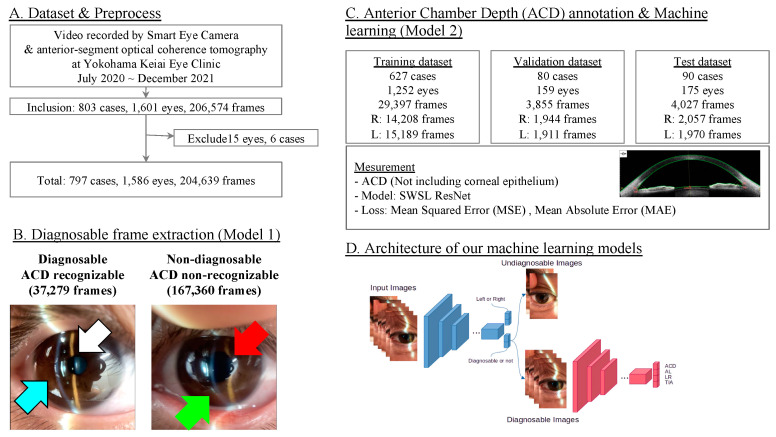
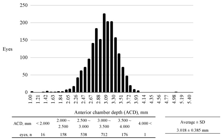
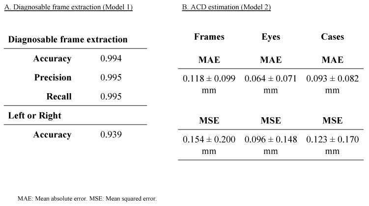
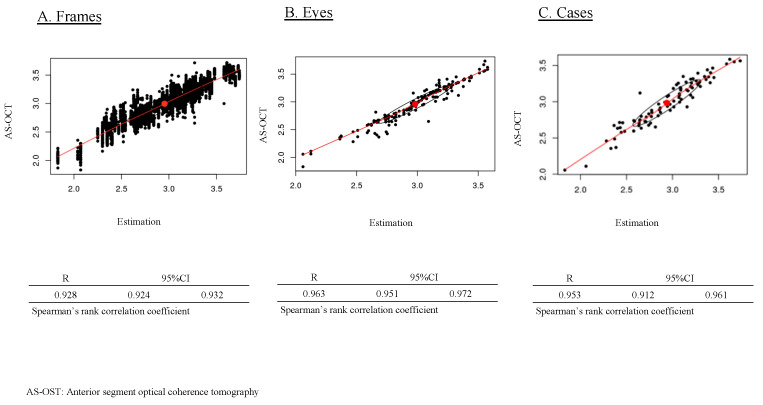
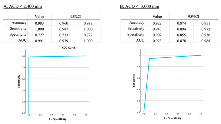
Primary angle closure glaucoma (PACG) is a major cause of visual impairment, particularly in Asia. Although effective screening tools are necessary, the current gold standard is complex and time-consuming, requiring extensive expertise. Artificial intelligence has introduced new opportunities for innovation in ophthalmic imaging. Anterior chamber depth (ACD) is a key risk factor for angle closure and has been suggested as a quick screening parameter for PACG. This study aims to develop an AI algorithm to quantitatively predict ACD from anterior segment photographs captured using a portable smartphone slit-lamp microscope. We retrospectively collected 204,639 frames from 1586 eyes, with ACD values obtained by anterior-segment OCT. We developed two models, (Model 1) diagnosable frame extraction and (Model 2) ACD estimation, using SWSL ResNet as the machine learning model. Model 1 achieved an accuracy of 0.994. Model 2 achieved an MAE of 0.093 ± 0.082 mm, an MSE of 0.123 ± 0.170 mm, and a correlation of R = 0.953. Furthermore, our model's estimation of the risk for angle closure showed a sensitivity of 0.943, specificity of 0.902, and an area under the curve (AUC) of 0.923 (95%CI: 0.878-0.968). We successfully developed a high-performance ACD estimation model, laying the groundwork for predicting other quantitative measurements relevant to PACG screening.

摘要

原发性闭角型青光眼(PACG)是导致视力损害的主要原因,在亚洲尤为如此。尽管有效的筛查工具必不可少,但目前的金标准复杂且耗时,需要广泛的专业知识。人工智能为眼科成像创新带来了新机遇。前房深度(ACD)是闭角的关键危险因素,已被提议作为PACG的快速筛查参数。本研究旨在开发一种人工智能算法,用于从使用便携式智能手机裂隙灯显微镜拍摄的眼前节照片中定量预测ACD。我们回顾性收集了来自1586只眼睛的204,639帧图像,通过眼前节OCT获得ACD值。我们使用SWSL ResNet作为机器学习模型开发了两个模型,(模型1)可诊断帧提取和(模型2)ACD估计。模型1的准确率达到0.994。模型2的平均绝对误差(MAE)为0.093±0.082毫米,均方误差(MSE)为0.123±0.170毫米,相关系数R = 0.953。此外,我们的模型对闭角风险的估计显示敏感性为0.943,特异性为0.902,曲线下面积(AUC)为0.923(95%CI:0.878 - 0.968)。我们成功开发了一个高性能的ACD估计模型,为预测与PACG筛查相关的其他定量测量奠定了基础。

https://cdn.ncbi.nlm.nih.gov/pmc/blobs/f6ec/11505230/025e1edb58f8/bioengineering-11-01005-g006.jpg
https://cdn.ncbi.nlm.nih.gov/pmc/blobs/f6ec/11505230/5f6dbabb14d2/bioengineering-11-01005-g001.jpg
https://cdn.ncbi.nlm.nih.gov/pmc/blobs/f6ec/11505230/ea137fdfffd4/bioengineering-11-01005-g002.jpg
https://cdn.ncbi.nlm.nih.gov/pmc/blobs/f6ec/11505230/7058c78157fc/bioengineering-11-01005-g003.jpg
https://cdn.ncbi.nlm.nih.gov/pmc/blobs/f6ec/11505230/435d476b3d83/bioengineering-11-01005-g004.jpg
https://cdn.ncbi.nlm.nih.gov/pmc/blobs/f6ec/11505230/9f7b71fce5bf/bioengineering-11-01005-g005.jpg
https://cdn.ncbi.nlm.nih.gov/pmc/blobs/f6ec/11505230/025e1edb58f8/bioengineering-11-01005-g006.jpg

相似文献

1
The Use of Artificial Intelligence for Estimating Anterior Chamber Depth from Slit-Lamp Images Developed Using Anterior-Segment Optical Coherence Tomography.利用人工智能从基于前段光学相干断层扫描技术生成的裂隙灯图像中估计前房深度
Bioengineering (Basel). 2024 Oct 9;11(10):1005. doi: 10.3390/bioengineering11101005.
2
From 2 dimensions to 3rd dimension: Quantitative prediction of anterior chamber depth from anterior segment photographs via deep-learning.从二维到三维:通过深度学习从眼前节照片定量预测前房深度
PLOS Digit Health. 2023 Feb 1;2(2):e0000193. doi: 10.1371/journal.pdig.0000193. eCollection 2023 Feb.
3
A Study Validating the Estimation of Anterior Chamber Depth and Iridocorneal Angle with Portable and Non-Portable Slit-Lamp Microscopy.一种利用便携与非便携裂隙灯显微镜对前房深度和房角进行评估的验证研究。
Sensors (Basel). 2021 Feb 19;21(4):1436. doi: 10.3390/s21041436.
4
Smartphone-Acquired Anterior Segment Images for Deep Learning Prediction of Anterior Chamber Depth: A Proof-of-Concept Study.用于前房深度深度学习预测的智能手机采集眼前节图像:一项概念验证研究。
Front Med (Lausanne). 2022 Jun 23;9:912214. doi: 10.3389/fmed.2022.912214. eCollection 2022.
5
Machine Learning-Guided Prediction of Central Anterior Chamber Depth Using Slit Lamp Images from a Portable Smartphone Device.基于便携智能手机裂隙灯图像的机器学习引导的中央前房深度预测
Biosensors (Basel). 2021 Jun 5;11(6):182. doi: 10.3390/bios11060182.
6
[Detection of the anterior segment configuration of primary angle-closure glaucoma by anterior segment optical coherence tomography].[眼前节光学相干断层扫描对原发性闭角型青光眼眼前节形态的检测]
Zhonghua Yan Ke Za Zhi. 2013 Feb;49(2):109-15.
7
Evaluation of white-to-white distance and anterior chamber depth measurements using the IOL Master, slit-lamp adapted optical coherence tomography and digital photographs in phakic eyes.使用IOL Master、裂隙灯适配光学相干断层扫描和数码照片评估有晶状体眼的白对白距离和前房深度测量值。
Klin Oczna. 2015;117(3):153-9.
8
Evaluation of the Diagnostic Performance of Swept-Source Anterior Segment Optical Coherence Tomography in Primary Angle Closure Disease.原发性闭角型青光眼的扫频源眼前节光学相干断层扫描诊断性能评估。
Am J Ophthalmol. 2022 Jan;233:68-77. doi: 10.1016/j.ajo.2021.06.033. Epub 2021 Jul 17.
9
Slit-lamp measurement of anterior chamber depth and its agreement with anterior segment optical coherence tomography and Lenstar LS 900 in pseudoexfoliation and normal eyes.前房深度的裂隙灯测量及其与前节光学相干断层扫描和 Lenstar LS 900 在假性剥脱和正常眼中的一致性。
Indian J Ophthalmol. 2021 Sep;69(9):2469-2474. doi: 10.4103/ijo.IJO_3738_20.
10
Non-contact tests for identifying people at risk of primary angle closure glaucoma.用于识别原发性闭角型青光眼高危人群的非接触式检测
Cochrane Database Syst Rev. 2020 May 28;5(5):CD012947. doi: 10.1002/14651858.CD012947.pub2.

引用本文的文献

1
Steroid-Responsive Intraocular Lens Deposits: A Case-Based Review of Diagnosis and Management.类固醇反应性人工晶状体沉积物:基于病例的诊断与管理综述
Cureus. 2025 Apr 23;17(4):e82878. doi: 10.7759/cureus.82878. eCollection 2025 Apr.
2
Keypoint localization and parameter measurement in ultrasound biomicroscopy anterior segment images based on deep learning.基于深度学习的超声生物显微镜眼前节图像中的关键点定位与参数测量
Biomed Eng Online. 2025 May 6;24(1):53. doi: 10.1186/s12938-025-01388-3.

本文引用的文献

1
Accuracy of Autonomous Artificial Intelligence-Based Diabetic Retinopathy Screening in Real-Life Clinical Practice.基于自主人工智能的糖尿病视网膜病变筛查在实际临床实践中的准确性
J Clin Med. 2024 Aug 14;13(16):4776. doi: 10.3390/jcm13164776.
2
A deep learning approach to explore the association of age-related macular degeneration polygenic risk score with retinal optical coherence tomography: A preliminary study.深度学习方法探索年龄相关性黄斑变性多基因风险评分与视网膜光学相干断层扫描的关联:一项初步研究。
Acta Ophthalmol. 2024 Nov;102(7):e1029-e1039. doi: 10.1111/aos.16710. Epub 2024 May 18.
3
Comparison of 21 artificial intelligence algorithms in automated diabetic retinopathy screening using handheld fundus camera.
比较使用手持眼底相机的 21 种人工智能算法在自动化糖尿病性视网膜病变筛查中的应用。
Ann Med. 2024 Dec;56(1):2352018. doi: 10.1080/07853890.2024.2352018. Epub 2024 May 13.
4
Transparency in Artificial Intelligence Reporting in Ophthalmology-A Scoping Review.眼科人工智能报告的透明度——一项范围综述
Ophthalmol Sci. 2024 Jan 18;4(4):100471. doi: 10.1016/j.xops.2024.100471. eCollection 2024 Jul-Aug.
5
Rate and Predictors of Misclassification of Active Diabetic Macular Edema as Detected by an Automated Retinal Image Analysis System.通过自动视网膜图像分析系统检测出的糖尿病性黄斑水肿误诊率及预测因素
Ophthalmol Ther. 2024 Jun;13(6):1553-1567. doi: 10.1007/s40123-024-00929-8. Epub 2024 Apr 8.
6
Development of the AI Pipeline for Corneal Opacity Detection.用于角膜混浊检测的人工智能流程开发。
Bioengineering (Basel). 2024 Mar 12;11(3):273. doi: 10.3390/bioengineering11030273.
7
Feasibility of Tear Meniscus Height Measurements Obtained with a Smartphone-Attachable Portable Device and Agreement of the Results with Standard Slit Lamp Examination.使用智能手机可连接便携式设备测量泪液半月板高度的可行性以及结果与标准裂隙灯检查的一致性。
Diagnostics (Basel). 2024 Feb 1;14(3):316. doi: 10.3390/diagnostics14030316.
8
Deep learning model for extensive smartphone-based diagnosis and triage of cataracts and multiple corneal diseases.基于深度学习的智能手机广泛诊断和分类白内障及多种角膜疾病的模型。
Br J Ophthalmol. 2024 Sep 20;108(10):1406-1413. doi: 10.1136/bjo-2023-324488.
9
AI-based diagnosis of nuclear cataract from slit-lamp videos.基于人工智能的裂隙灯视频核性白内障诊断。
Sci Rep. 2023 Dec 12;13(1):22046. doi: 10.1038/s41598-023-49563-7.
10
EyeArt artificial intelligence analysis of diabetic retinopathy in retinal screening events.视网膜筛查中EyeArt人工智能对糖尿病视网膜病变的分析
Int Ophthalmol. 2023 Dec;43(12):4851-4859. doi: 10.1007/s10792-023-02887-9. Epub 2023 Oct 17.