• 文献检索
  • 文档翻译
  • 深度研究
  • 学术资讯
  • Suppr Zotero 插件Zotero 插件
  • 邀请有礼
  • 套餐&价格
  • 历史记录
应用&插件
Suppr Zotero 插件Zotero 插件浏览器插件Mac 客户端Windows 客户端微信小程序
定价
高级版会员购买积分包购买API积分包
服务
文献检索文档翻译深度研究API 文档MCP 服务
关于我们
关于 Suppr公司介绍联系我们用户协议隐私条款
关注我们

Suppr 超能文献

核心技术专利:CN118964589B侵权必究
粤ICP备2023148730 号-1Suppr @ 2026

文献检索

告别复杂PubMed语法,用中文像聊天一样搜索,搜遍4000万医学文献。AI智能推荐,让科研检索更轻松。

立即免费搜索

文件翻译

保留排版,准确专业,支持PDF/Word/PPT等文件格式,支持 12+语言互译。

免费翻译文档

深度研究

AI帮你快速写综述,25分钟生成高质量综述,智能提取关键信息,辅助科研写作。

立即免费体验

新型冠状病毒肺炎的冠状动脉计算机断层扫描血管造影(CTA)表现

Coronary Computed Tomography Angiography (CTA) Findings in COVID-19.

作者信息

Lacaita Pietro G, Luger Anna, Plank Fabian, Barbieri Fabian, Beyer Christoph, Thurner Theresa, Scharll Yannick, Deeg Johannes, Widmann Gerlig, Feuchtner Gudrun M

机构信息

Department Radiology, Innsbruck Medical University, 6020 Innsbruck, Austria.

Department Internal Medicine, Tyrol Clinicum Hall, 6060 Hall, Austria.

出版信息

J Cardiovasc Dev Dis. 2024 Oct 14;11(10):325. doi: 10.3390/jcdd11100325.

DOI:10.3390/jcdd11100325
PMID:39452295
原文链接:https://pmc.ncbi.nlm.nih.gov/articles/PMC11508304/
Abstract

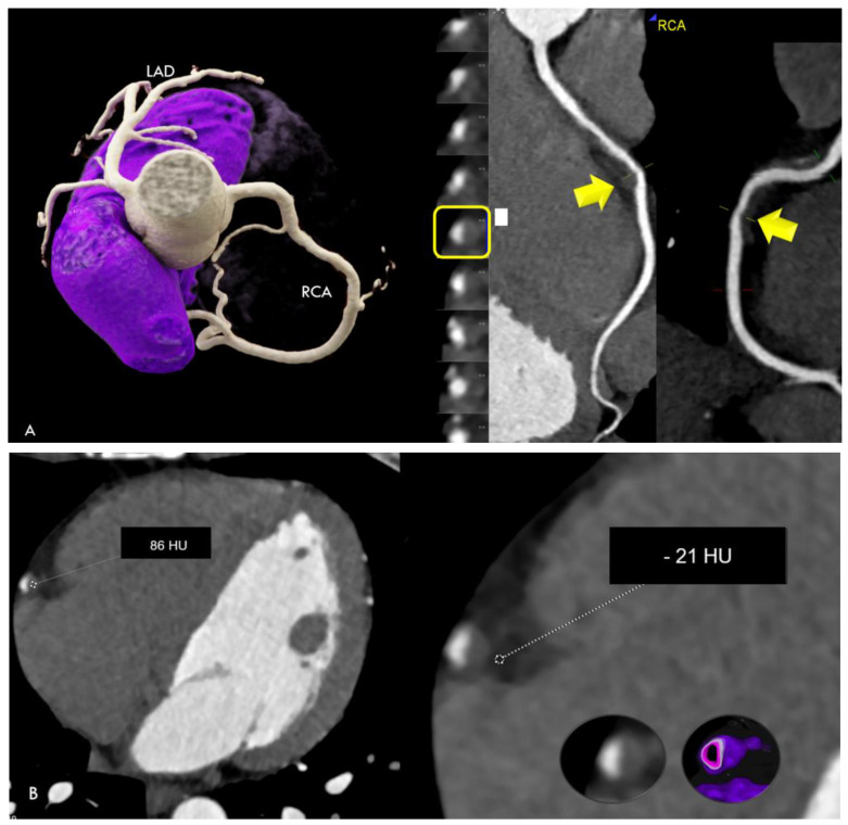
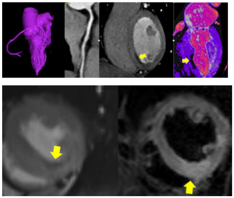
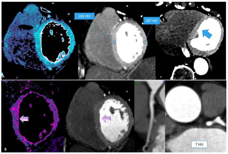
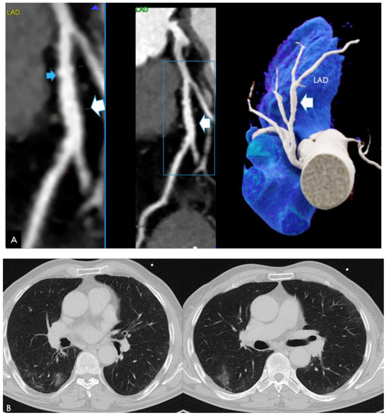
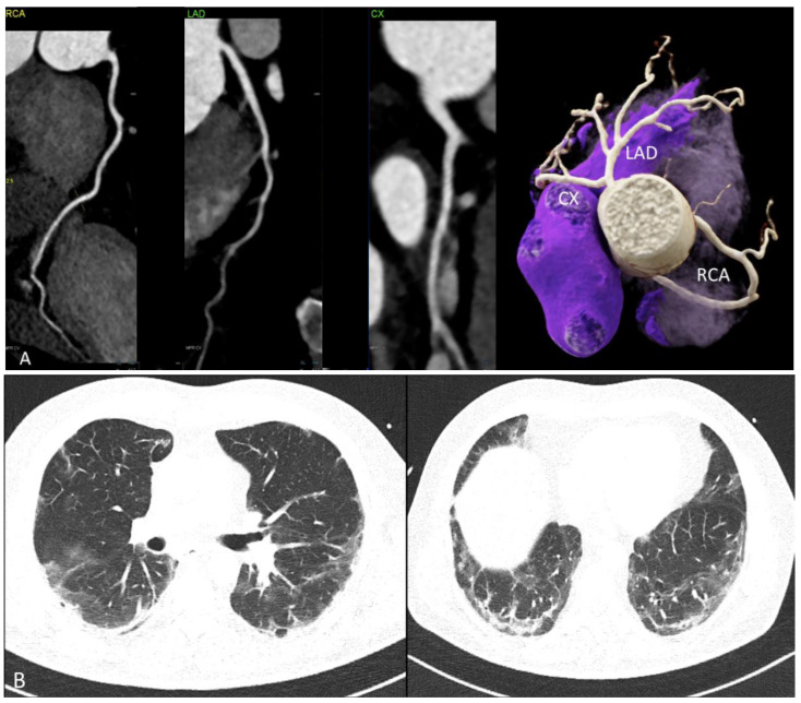
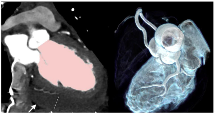
(1) Background: The novel SARS-CoV-2 virus infects the endothelium. Vasculitis may lead to specific coronary artery wall lesions. Coronary computed tomography angiography (CTA) imaging findings have not been systematically reported. The aim of this study was to describe a case series using CTA. (2) Methods: Patients with recent RT-PCR confirmed SARS-CoV-2 infection referred for coronary CTA for clinical indications (e.g., chest pain, troponin+, and ECG abnormalities) were included. Coronary CTA findings, such as atypical coronary lesions suggestive of vasculitis, perivascular inflammation measured by using pericoronary fat attenuation (PCAT) index, coronary artery disease, and extracoronary findings were collected. (3) Results: Results for 12 patients (54.8 ± 22 years; four females) with SARS-CoV-2 infection within 60 days (four acute care and eight stable patients) are reported. Time to positive RT-PCR was a mean of 15.1 days (range, 0-51). In four acute patients with signs of myocardial injury, plaque rupture (n = 1), hyperenhancing myocardium/MINOCA (n = 1), MINOCA (n = 1), and pericarditis with acute heart failure (LVEF 20%) (n = 1) were found. All (100%) had pericardial effusion and signs of perivascular inflammation. Among eight stable patients, pericardial effusion or perivascular inflammation were found in only two (25%). Coronary artery disease was ruled out in five (62.5%) (4) Conclusions: Coronary CTA is a useful imaging modality in the diagnostic work up of patients with COVID-19 infection, and is able to describe coronary and other cardiac abnormalities.

摘要

(1) 背景:新型严重急性呼吸综合征冠状病毒2(SARS-CoV-2)可感染内皮细胞。血管炎可能导致特定的冠状动脉壁病变。冠状动脉计算机断层扫描血管造影(CTA)成像结果尚未得到系统报道。本研究旨在描述一组使用CTA的病例系列。(2) 方法:纳入近期经逆转录聚合酶链反应(RT-PCR)确诊为SARS-CoV-2感染、因临床指征(如胸痛、肌钙蛋白阳性和心电图异常)而接受冠状动脉CTA检查的患者。收集冠状动脉CTA检查结果,如提示血管炎的非典型冠状动脉病变、使用冠状动脉周围脂肪衰减(PCAT)指数测量的血管周围炎症、冠状动脉疾病及冠状动脉外检查结果。(3) 结果:报告了12例在60天内感染SARS-CoV-2的患者(年龄54.8±22岁;4名女性)(4例急性护理患者和8例稳定患者)。RT-PCR检测呈阳性的平均时间为15.1天(范围0-51天)。在4例有心肌损伤迹象的急性患者中,发现斑块破裂(n = 1)、心肌强化/无阻塞性冠状动脉粥样硬化心脏病(MINOCA)(n = 1)、MINOCA(n = 1)以及伴有急性心力衰竭(左心室射血分数20%)的心包炎(n = 1)。所有患者(100%)均有心包积液和血管周围炎症迹象。在8例稳定患者中,仅2例(25%)发现心包积液或血管周围炎症。5例(62.5%)排除了冠状动脉疾病。(4) 结论:冠状动脉CTA是COVID-19感染患者诊断检查中的一种有用成像方式,能够描述冠状动脉及其他心脏异常情况。

https://cdn.ncbi.nlm.nih.gov/pmc/blobs/ce16/11508304/ffc3fa669b42/jcdd-11-00325-g006.jpg
https://cdn.ncbi.nlm.nih.gov/pmc/blobs/ce16/11508304/8041ccaeb862/jcdd-11-00325-g001a.jpg
https://cdn.ncbi.nlm.nih.gov/pmc/blobs/ce16/11508304/45ff65de7946/jcdd-11-00325-g002.jpg
https://cdn.ncbi.nlm.nih.gov/pmc/blobs/ce16/11508304/c05834d4b2d6/jcdd-11-00325-g003.jpg
https://cdn.ncbi.nlm.nih.gov/pmc/blobs/ce16/11508304/dbeeb0fdbc52/jcdd-11-00325-g004.jpg
https://cdn.ncbi.nlm.nih.gov/pmc/blobs/ce16/11508304/6183758aad71/jcdd-11-00325-g005.jpg
https://cdn.ncbi.nlm.nih.gov/pmc/blobs/ce16/11508304/ffc3fa669b42/jcdd-11-00325-g006.jpg

相似文献

1
Coronary Computed Tomography Angiography (CTA) Findings in COVID-19.新型冠状病毒肺炎的冠状动脉计算机断层扫描血管造影(CTA)表现
J Cardiovasc Dev Dis. 2024 Oct 14;11(10):325. doi: 10.3390/jcdd11100325.
2
High pericoronary adipose tissue attenuation on computed tomography angiography predicts cardiovascular events in patients with type 2 diabetes mellitus: post-hoc analysis from a prospective cohort study.冠状动脉周围脂肪组织 CT 血管造影衰减程度高可预测 2 型糖尿病患者的心血管事件:前瞻性队列研究的事后分析。
Cardiovasc Diabetol. 2022 Mar 18;21(1):44. doi: 10.1186/s12933-022-01478-9.
3
Pericoronary Adipose Tissue Computed Tomography Attenuation and High-Risk Plaque Characteristics in Acute Coronary Syndrome Compared With Stable Coronary Artery Disease.对比稳定型冠心病,急性冠状动脉综合征患者的冠状动脉旁脂肪组织 CT 衰减值与高危斑块特征。
JAMA Cardiol. 2018 Sep 1;3(9):858-863. doi: 10.1001/jamacardio.2018.1997.
4
Value of Myocardial Perfusion Assessment With Coronary Computed Tomography Angiography in Patients With Recent Acute-Onset Chest Pain.近期急性发作胸痛患者冠状动脉计算机断层扫描血管造影心肌灌注评估的价值。
JACC Cardiovasc Imaging. 2018 Nov;11(11):1611-1621. doi: 10.1016/j.jcmg.2017.09.022. Epub 2017 Dec 13.
5
Coronary Inflammation by Computed Tomography Pericoronary Fat Attenuation in MINOCA and Tako-Tsubo Syndrome.MINOCA 和 Takotsubo 综合征患者冠状动脉 CT 扫描心包脂肪衰减与冠状动脉炎症。
J Am Heart Assoc. 2019 Sep 3;8(17):e013235. doi: 10.1161/JAHA.119.013235. Epub 2019 Aug 29.
6
Association between High Pericoronary Adipose Tissue Computed Tomography Attenuation and Impaired Flow-Mediated Dilation of the Brachial Artery.冠状动脉周围脂肪组织 CT 衰减值与肱动脉血流介导的舒张功能障碍的相关性。
J Atheroscler Thromb. 2023 Apr 1;30(4):364-376. doi: 10.5551/jat.63580. Epub 2022 Jun 24.
7
Relationship between changes in pericoronary adipose tissue attenuation and coronary plaque burden quantified from coronary computed tomography angiography.基于冠状动脉 CT 血管造影定量评估的冠状动脉斑块负荷与冠状动脉脂肪衰减变化的关系。
Eur Heart J Cardiovasc Imaging. 2019 Jun 1;20(6):636-643. doi: 10.1093/ehjci/jez013.
8
Non-invasive detection of coronary inflammation using computed tomography and prediction of residual cardiovascular risk (the CRISP CT study): a post-hoc analysis of prospective outcome data.使用计算机断层扫描技术无创性检测冠状动脉炎症及预测残余心血管风险(CRISP CT 研究):前瞻性结局数据的事后分析。
Lancet. 2018 Sep 15;392(10151):929-939. doi: 10.1016/S0140-6736(18)31114-0. Epub 2018 Aug 28.
9
Assessing the Impact of Long-Term High-Dose Statin Treatment on Pericoronary Inflammation and Plaque Distribution-A Comprehensive Coronary CTA Follow-Up Study.评估长期大剂量他汀类药物治疗对冠状动脉周围炎症和斑块分布的影响——一项综合冠状动脉 CTA 随访研究。
Int J Mol Sci. 2024 Jan 30;25(3):1700. doi: 10.3390/ijms25031700.
10
Pericoronary Adipose Tissue Attenuation in Patients With Acute Coronary Syndrome Versus Stable Coronary Artery Disease.急性冠状动脉综合征与稳定型冠状动脉疾病患者的冠状动脉旁脂肪组织衰减。
Circ Cardiovasc Imaging. 2023 Feb;16(2):e014672. doi: 10.1161/CIRCIMAGING.122.014672. Epub 2023 Feb 3.

本文引用的文献

1
Charting the Unseen: How Non-Invasive Imaging Could Redefine Cardiovascular Prevention.绘制不可见之物:无创成像如何重新定义心血管疾病预防
J Cardiovasc Dev Dis. 2024 Aug 9;11(8):245. doi: 10.3390/jcdd11080245.
2
Photon-Counting Detector Computed Tomography (PCD-CT): A New Era for Cardiovascular Imaging? Current Status and Future Outlooks.光子计数探测器计算机断层扫描(PCD-CT):心血管成像的新时代?现状与未来展望。
J Cardiovasc Dev Dis. 2024 Apr 21;11(4):127. doi: 10.3390/jcdd11040127.
3
Characteristics of patients with recurrent acute myocardial infarction after MINOCA.
MINOCA 后复发性急性心肌梗死患者的特征。
Prog Cardiovasc Dis. 2023 Nov-Dec;81:42-47. doi: 10.1016/j.pcad.2023.10.006. Epub 2023 Oct 16.
4
Myocardial extracellular volume quantification with computed tomography-current status and future outlook.基于计算机断层扫描的心肌细胞外容积定量——现状与未来展望
Insights Imaging. 2023 Sep 25;14(1):156. doi: 10.1186/s13244-023-01506-6.
5
Computed tomography coronary angiography in non-ST-segment elevation myocardial infarction.非 ST 段抬高型心肌梗死的计算机断层冠状动脉成像。
Br J Radiol. 2022 Dec 1;95(1140):20220346. doi: 10.1259/bjr.20220346. Epub 2022 Sep 20.
6
Cardiometabolic outcomes up to 12 months after COVID-19 infection. A matched cohort study in the UK.感染新冠病毒后 12 个月内的心脏代谢结局。英国一项匹配队列研究。
PLoS Med. 2022 Jul 19;19(7):e1004052. doi: 10.1371/journal.pmed.1004052. eCollection 2022 Jul.
7
Preventive cardiology advances in the 2021 AHA/ACC chest pain guideline.2021年美国心脏协会/美国心脏病学会胸痛指南中的预防心脏病学进展。
Am J Prev Cardiol. 2022 Jun 30;11:100365. doi: 10.1016/j.ajpc.2022.100365. eCollection 2022 Sep.
8
Cardiovascular outcome 6 months after severe coronavirus disease 2019 infection.严重新型冠状病毒 2019 感染后 6 个月的心血管结局。
J Hypertens. 2022 Jul 1;40(7):1278-1287. doi: 10.1097/HJH.0000000000003110. Epub 2022 Feb 25.
9
Extracellular Volume Quantification With Cardiac Late Enhancement Scanning Using Dual-Source Photon-Counting Detector CT.应用双源光子计数探测器 CT 行心脏晚期钆增强扫描对细胞外容积进行定量评估。
Invest Radiol. 2022 Jun 1;57(6):406-411. doi: 10.1097/RLI.0000000000000851. Epub 2022 Jan 21.
10
2021 AHA/ACC/ASE/CHEST/SAEM/SCCT/SCMR Guideline for the Evaluation and Diagnosis of Chest Pain: A Report of the American College of Cardiology/American Heart Association Joint Committee on Clinical Practice Guidelines.2021 AHA/ACC/ASE/CHEST/SAEM/SCCT/SCMR 胸痛评估与诊断指南:美国心脏病学会/美国心脏协会联合临床实践指南委员会的报告。
Circulation. 2021 Nov 30;144(22):e368-e454. doi: 10.1161/CIR.0000000000001029. Epub 2021 Oct 28.