Department of Molecular Life Sciences, University of Zurich (UZH), Zurich, Switzerland.
Center for Microscopy and Image Analyses, University of Zurich (UZH), Zurich, Switzerland.
Sci Adv. 2024 Oct 25;10(43):eadq7483. doi: 10.1126/sciadv.adq7483.
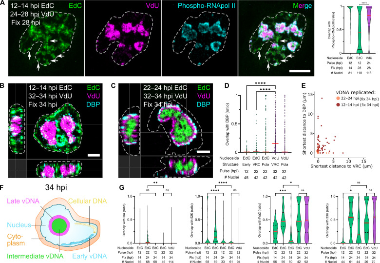
Biomolecular assemblies are fundamental to life and viral disease. The spatiotemporal coordination of viral replication and assembly is largely unknown. Here, we developed a dual-color click chemistry procedure for imaging adenovirus DNA (vDNA) replication in the cell nucleus. Late- but not early-replicated vDNA was packaged into virions. Early-replicated vDNA segregated from the viral replication compartment (VRC). Single object tracking, superresolution microscopy, fluorescence recovery after photobleaching, and correlative light-electron microscopy revealed a stepwise assembly program involving vDNA and capsid intermediates. Depending on replication and the scaffolding protein 52K, late-replicated vDNA with rapidly exchanging green fluorescent protein-tagged capsid linchpin protein V and incomplete virions emerged from the VRC periphery. These nanogel-like puncta exhibited restricted movements and were located with the capsid proteins hexon, VI, and virions in the nuclear periphery, suggestive of sites for virion formation. Our findings identify VRC dynamics and assembly intermediates, essential for stepwise productive adenovirus morphogenesis.
生物分子组装是生命和病毒疾病的基础。病毒复制和组装的时空协调在很大程度上是未知的。在这里,我们开发了一种双色点击化学程序,用于在细胞核中成像腺病毒 DNA(vDNA)复制。晚期而非早期复制的 vDNA 被包装成病毒粒子。早期复制的 vDNA 与病毒复制区室(VRC)分离。单个物体跟踪、超分辨率显微镜、光漂白后荧光恢复和相关的光电子显微镜揭示了一个涉及 vDNA 和衣壳中间体的逐步组装程序。根据复制和支架蛋白 52K,晚期复制的 vDNA 与快速交换绿色荧光蛋白标记的衣壳销蛋白 V 和不完全的病毒粒子从 VRC 外围出现。这些纳米凝胶样小点表现出受限的运动,并与衣壳蛋白六邻体、VI 和病毒粒子一起位于核周,提示病毒形成的部位。我们的发现确定了 VRC 动力学和组装中间体,这对逐步产生的腺病毒形态发生至关重要。