IRCCS Ospedale Policlinico San Martino, Largo Rosanna Benzi 10, 16132 Genova, Italy.
Department of Neurosciences, Rehabilitation, Ophthalmology, Genetics, and Maternal and Children's Sciences (DINOGMI), University of Genoa, 16126 Genova, Italy.
Int J Mol Sci. 2024 Oct 19;25(20):11244. doi: 10.3390/ijms252011244.
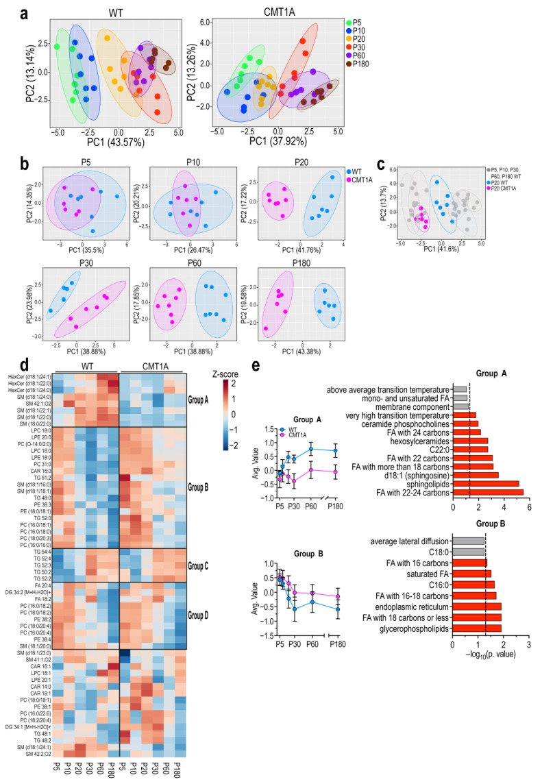
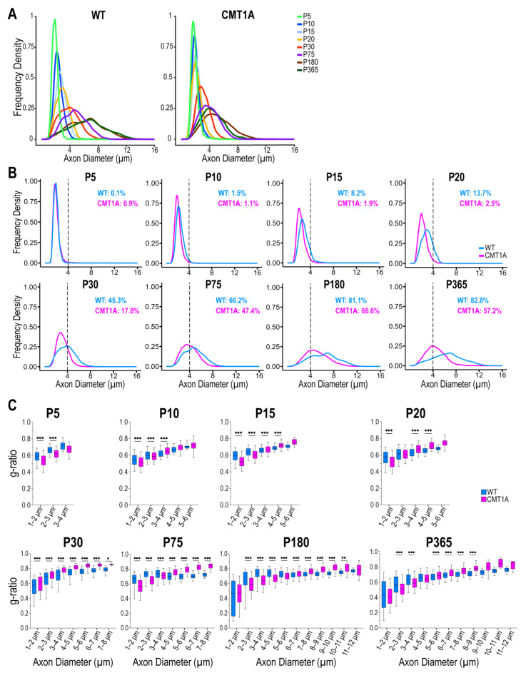
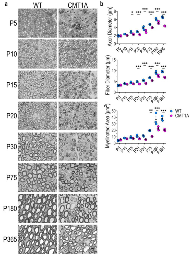
Findings accumulated over time show that neurophysiological, neuropathological, and molecular alterations are present in CMT1A and support the dysmyelinating rather than demyelinating nature of this neuropathy. Moreover, uniform slowing of nerve conduction velocity is already manifest in CMT1A children and does not improve throughout their life. This evidence and our previous studies displaying aberrant myelin composition and structure in adult CMT1A rats prompt us to hypothesize a myelin and axon developmental defect in the CMT1A peripheral nervous system. Peripheral myelination begins during the early stages of development in mammals and, during this process, chemical and structural features of myelinated fibers (MFs) evolve towards a mature phenotype; deficiencies within this self-modulating circuit can cause its blockage. Therefore, to shed light on pathophysiological mechanisms that occur during development, and to investigate the relationship among axonal, myelin, and lipidome deficiencies in CMT1A, we extensively analyzed the evolution of both myelin lipid profile and MF structure in WT and CMT1A rats. Lipidomic analysis revealed a delayed maturation of CMT1A myelin already detectable at P10 characterized by a deprivation of sphingolipid species such as hexosylceramides and long-chain sphingomyelins, whose concentration physiologically increases in WT, and an increase in lipids typical of unspecialized plasma membranes, including phosphatidylcholines and phosphatidylethanolamines. Consistently, advanced morphometric analysis on more than 130,000 MFs revealed a delay in the evolution of CMT1A axon and myelin geometric parameters, appearing concomitantly with lipid impairment. We here demonstrate that, during normal development, MFs undergo a continuous maturation process in both chemical composition and physical structure, but these processes are delayed in CMT1A.
随着时间的推移,研究结果表明,CMT1A 存在神经生理学、神经病理学和分子改变,支持这种神经病变为脱髓鞘而不是脱髓鞘的性质。此外,CMT1A 患儿的神经传导速度均匀减慢,且在其整个生命周期中均未改善。这一证据以及我们之前的研究显示,CMT1A 大鼠的异常髓鞘组成和结构促使我们假设 CMT1A 周围神经系统存在髓鞘和轴突发育缺陷。哺乳动物的周围髓鞘形成始于早期发育阶段,在此过程中,髓鞘纤维(MFs)的化学和结构特征向成熟表型发展;该自我调节回路中的缺陷会导致其阻塞。因此,为了阐明发育过程中发生的病理生理机制,并研究 CMT1A 中轴突、髓鞘和脂质组之间的关系,我们广泛分析了 WT 和 CMT1A 大鼠的髓鞘脂质谱和 MF 结构的演变。脂质组学分析显示,CMT1A 髓鞘的成熟延迟,在 P10 时即可检测到,其特征是鞘脂如己糖神经酰胺和长链神经鞘氨醇的缺乏,而 WT 中的这些物质的浓度会生理性增加,以及包括磷脂酰胆碱和磷脂乙醇胺在内的非特化质膜的脂质增加。一致地,对超过 130000 个 MF 进行的高级形态计量学分析显示,CMT1A 轴突和髓鞘几何参数的演变延迟,与脂质损伤同时出现。我们在这里证明,在正常发育过程中,MFs 在化学组成和物理结构上都经历了一个连续的成熟过程,但这些过程在 CMT1A 中延迟了。