Department of Neurology and Institute of Neurology, Ruijin Hospital, Shanghai Jiao Tong University School of Medicine, Shanghai, China.
Department of Ophthalmology, Ruijin Hospital, Shanghai Jiao Tong University School of Medicine, Shanghai, China.
PLoS One. 2024 Oct 31;19(10):e0312534. doi: 10.1371/journal.pone.0312534. eCollection 2024.
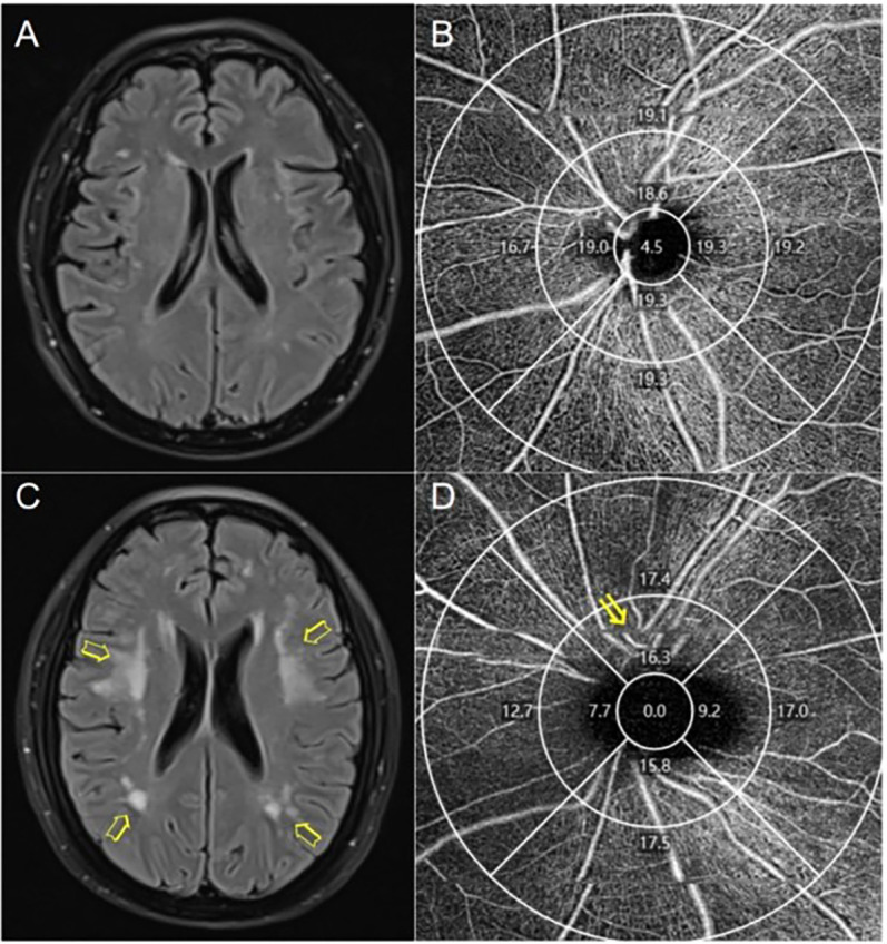
Chronic cerebral hypoperfusion triggers the development of white matter hyperintensities (WMHs), common in cerebral small vessel disease (CSVD). However, conventional imaging techniques cannot visualize cerebral small vessels. The retina, a direct extension of the central nervous system, has an unclear correlation with WMHs. This study employs Optical coherence tomographic angiography (OCTA) to investigate vascular changes in the retina and explore its correlation with WMHs, aiming to provide a new method for assessing perfusion in early ischemic brain WMHs.
Forty-nine patients with WMHs were stratified into mild and moderate/severe WMHs groups based on MRI findings, utilizing the Fazekas and Scheltens scales. OCTA assessed fundus vessel microcirculation. Logistic regression analyzed the correlation between ocular fundus microcirculation and WMH severity and location. Additionally, ROC curves evaluated the diagnostic efficacy of each fundus vascular microcirculation index in determining WMH severity.
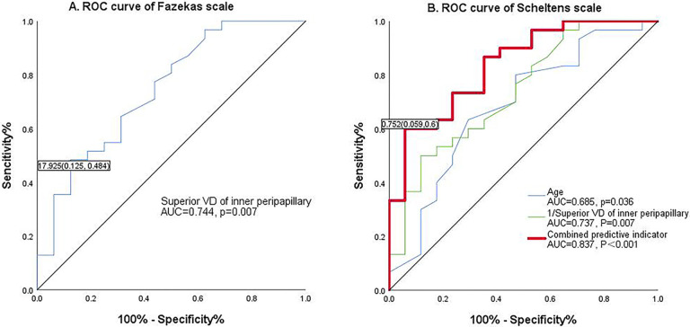
After adjusting for multiple confounders, finding consistently indicated that the moderate/ severe WMHs group exhibited lower vessel density (VD) in the superior quadrant of the inner peripapillary region compared to the mild group [OR = 0.487, CI (0.255,0.929), p < 0.05]. ROC curves revealed that when combined with age, diabetes, and superior quadrant VD of the inner peripapillary region, specificity could be increased to 94.1%.
Peripapillary vessel density correlates closely with the severity of cerebral WMHs. Early morphological changes due to chronic hypoperfusion may initiate from the inner layer of the optic disc, and OCTA could offer a novel method for evaluating blood perfusion in ischemic WMHs.
慢性脑灌注不足会引发脑白质高信号(WMHs)的发展,这在脑小血管病(CSVD)中很常见。然而,常规的成像技术无法观察到脑小血管。视网膜是中枢神经系统的直接延伸,与 WMHs 的相关性尚不清楚。本研究采用光相干断层扫描血管造影(OCTA)来研究视网膜血管的变化,并探讨其与 WMHs 的相关性,旨在为评估早期缺血性脑 WMHs 的灌注提供一种新方法。
根据 MRI 结果,将 49 例 WMHs 患者分为轻度和中重度/重度 WMHs 组,使用 Fazekas 和 Scheltens 量表。OCTA 评估眼底血管微循环。Logistic 回归分析眼底部微循环与 WMH 严重程度和位置的相关性。此外,ROC 曲线评估每个眼底血管微循环指数在确定 WMH 严重程度方面的诊断效能。
在调整了多个混杂因素后,研究结果一致表明,与轻度组相比,中重度/重度 WMHs 组内直肌旁视盘内上象限的血管密度(VD)较低[OR = 0.487,CI(0.255,0.929),p < 0.05]。ROC 曲线显示,当与年龄、糖尿病和内直肌旁视盘内上象限的 VD 相结合时,特异性可提高至 94.1%。
视盘旁血管密度与脑 WMHs 的严重程度密切相关。慢性灌注不足引起的早期形态学变化可能始于视盘内层,OCTA 可能为评估缺血性 WMHs 的血液灌注提供一种新方法。