Fu Wang, Zhou Xiaoyu, Wang Minli, Li Ping, Hou Jingjing, Gao Peng, Wang Jue
Department of Neurology, Shanghai Tenth People's Hospital, Tongji University School of Medicine, Shanghai, China.
Department of Ophthalmology, Shanghai Tenth People's Hospital, Tongji University School of Medicine, Shanghai, China.
Front Neurol. 2022 Apr 25;13:843198. doi: 10.3389/fneur.2022.843198. eCollection 2022.
To detect fundus changes in patients with cerebral small vessel disease (CSVD) using optical coherence tomography angiography (OCTA) and to investigate the correlations between CSVD and fundus changes.
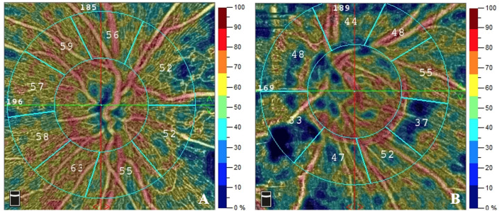
From January 2019 to January 2020, patients diagnosed with CSVD by magnetic resonance imaging (MRI) were enrolled in our study and received fundus examinations using OCTA. CSVD was defined as white matter hyperintensities, enlarged perivascular spaces, lacunes, or microbleeds on MRI. OCTA parameters included foveal avascular zone areas, retinal nerve fiber layer thickness, and capillary densities of the superficial retinal capillary plexuses, deep retinal capillary plexuses, and the radial peripapillary capillary network of the disc. Univariate and multivariate logistic regression analyses were performed to explore the correlation between CSVD and fundus changes.
A total of 115 patients (40% male) were enrolled and analyzed, and the mean age was 65.11 ± 11.23 years. After multivariate logistic regression analysis, the radial peripapillary capillary network density was negatively correlated with severity of deep white matter lesions (OR: 0.909; 95% CI: 0.828-0.998; = 0.046) and perivascular spaces (OR: 0.881; 95% CI: 0.779-0.995; = 0.041). Parafoveal vessel densities of the superficial retinal capillary plexuses were independently correlated with lacunes (OR: 0.889; 95% CI: 0.817-0.967; = 0.006).
OCTA parameters were correlated with CSVD, indicating that OCTA is a potential method for CSVD screening.
利用光学相干断层扫描血管造影(OCTA)检测脑小血管病(CSVD)患者的眼底变化,并探讨CSVD与眼底变化之间的相关性。
2019年1月至2020年1月,将通过磁共振成像(MRI)诊断为CSVD的患者纳入本研究,并使用OCTA进行眼底检查。CSVD定义为MRI上的白质高信号、血管周围间隙扩大、腔隙或微出血。OCTA参数包括中心凹无血管区面积、视网膜神经纤维层厚度,以及浅表视网膜毛细血管丛、深部视网膜毛细血管丛和视盘周围放射状毛细血管网的毛细血管密度。进行单因素和多因素逻辑回归分析,以探讨CSVD与眼底变化之间的相关性。
共纳入并分析了115例患者(40%为男性),平均年龄为65.11±11.23岁。多因素逻辑回归分析后,视盘周围放射状毛细血管网密度与深部白质病变严重程度呈负相关(OR:0.909;95%CI:0.828 - 0.998;P = 0.046),与血管周围间隙呈负相关(OR:0.881;95%CI:0.779 - 0.995;P = 0.041)。浅表视网膜毛细血管丛的黄斑旁血管密度与腔隙独立相关(OR:0.889;95%CI:0.817 - 0.967;P = 0.006)。
OCTA参数与CSVD相关,表明OCTA是一种用于CSVD筛查的潜在方法。