• 文献检索
  • 文档翻译
  • 深度研究
  • 学术资讯
  • Suppr Zotero 插件Zotero 插件
  • 邀请有礼
  • 套餐&价格
  • 历史记录
应用&插件
Suppr Zotero 插件Zotero 插件浏览器插件Mac 客户端Windows 客户端微信小程序
定价
高级版会员购买积分包购买API积分包
服务
文献检索文档翻译深度研究API 文档MCP 服务
关于我们
关于 Suppr公司介绍联系我们用户协议隐私条款
关注我们

Suppr 超能文献

核心技术专利:CN118964589B侵权必究
粤ICP备2023148730 号-1Suppr @ 2026

文献检索

告别复杂PubMed语法,用中文像聊天一样搜索,搜遍4000万医学文献。AI智能推荐,让科研检索更轻松。

立即免费搜索

文件翻译

保留排版,准确专业,支持PDF/Word/PPT等文件格式,支持 12+语言互译。

免费翻译文档

深度研究

AI帮你快速写综述,25分钟生成高质量综述,智能提取关键信息,辅助科研写作。

立即免费体验

正常衰老成年人视网膜微血管与脑衰弱的影像标志物

Retinal microvasculature and imaging markers of brain frailty in normal aging adults.

作者信息

Tao Wendan, Kwapong William Robert, Xie Jianyang, Wang Zetao, Guo Xiaonan, Liu Junfeng, Ye Chen, Wu Bo, Zhao Yitian, Liu Ming

机构信息

Department of Neurology, West China Hospital, Sichuan University, Chengdu, China.

Cixi Institute of Biomedical Engineering, Ningbo Institute of Materials Technology and Engineering, Chinese Academy of Sciences, Ningbo, China.

出版信息

Front Aging Neurosci. 2022 Aug 22;14:945964. doi: 10.3389/fnagi.2022.945964. eCollection 2022.

DOI:10.3389/fnagi.2022.945964
PMID:36072485
原文链接:https://pmc.ncbi.nlm.nih.gov/articles/PMC9441884/
Abstract

BACKGROUND

The retina and brain share a similar embryologic origin, blood barriers, and microvasculature features. Thus, retinal imaging has been of interest in the aging population to help in the early detection of brain disorders. Imaging evaluation of brain frailty, including brain atrophy and markers of cerebral small vessel disease (CSVD), could reflect brain health in normal aging, but is costly and time-consuming. In this study, we aimed to evaluate the retinal microvasculature and its association with radiological indicators of brain frailty in normal aging adults.

METHODS

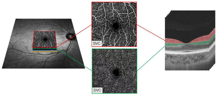
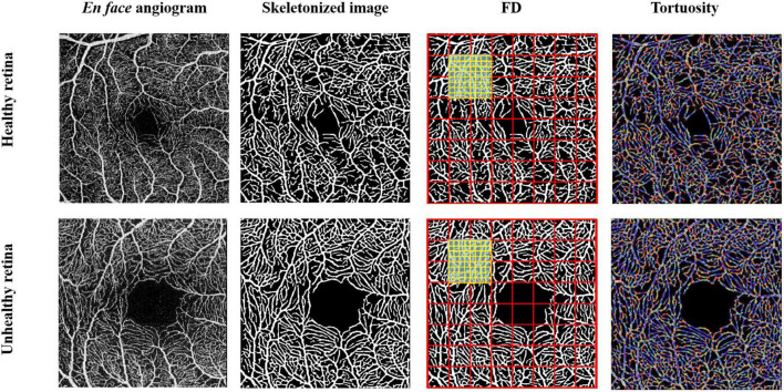
Swept-source optical coherence tomography angiography (SS-OCTA) and 3T-MRI brain scanning were performed on normal aging adults (aged ≥ 50 years). Using a deep learning algorithm, microvascular tortuosity (VT) and fractal dimension parameter (D) were used to evaluate the superficial vascular complex (SVC) and deep vascular complex (DVC) of the retina. MRI markers of brain frailty include brain volumetric measures and CSVD markers that were assessed.

RESULTS

Of the 139 normal aging individuals included, the mean age was 59.43 ± 7.31 years, and 64.0% ( = 89) of the participants were females. After adjustment of age, sex, and vascular risk factors, D in the DVC showed a significant association with the presence of lacunes (β = 0.58, = 0.007), while VT in the SVC significantly correlated with the score of cerebral deep white matter hyperintensity (β = 0.31, = 0.027). No correlations were found between brain volumes and retinal microvasculature changes ( > 0.05).

CONCLUSION

Our report suggests that imaging of the retinal microvasculature may give clues to brain frailty in the aging population.

摘要

背景

视网膜和大脑具有相似的胚胎起源、血脑屏障和微血管特征。因此,视网膜成像在老年人群中受到关注,有助于早期发现脑部疾病。对脑衰弱的成像评估,包括脑萎缩和脑小血管疾病(CSVD)的标志物,可反映正常衰老过程中的脑健康状况,但成本高且耗时。在本研究中,我们旨在评估正常衰老成年人的视网膜微血管及其与脑衰弱放射学指标的关联。

方法

对正常衰老成年人(年龄≥50岁)进行扫频光学相干断层扫描血管造影(SS-OCTA)和3T磁共振成像(MRI)脑部扫描。使用深度学习算法,微血管迂曲度(VT)和分形维数参数(D)用于评估视网膜的浅表血管复合体(SVC)和深部血管复合体(DVC)。评估脑衰弱的MRI标志物包括脑容量测量和CSVD标志物。

结果

纳入的139名正常衰老个体中,平均年龄为59.43±7.31岁,64.0%(n = 89)的参与者为女性。在调整年龄、性别和血管危险因素后,DVC中的D与腔隙的存在显著相关(β = 0.58,P = 0.007),而SVC中的VT与脑深部白质高信号评分显著相关(β = 0.31,P = 0.027)。未发现脑容量与视网膜微血管变化之间存在相关性(P>0.05)。

结论

我们的报告表明视网膜微血管成像可能为老年人群的脑衰弱提供线索。

https://cdn.ncbi.nlm.nih.gov/pmc/blobs/53c7/9441884/6e7f9d8b58e1/fnagi-14-945964-g002.jpg
https://cdn.ncbi.nlm.nih.gov/pmc/blobs/53c7/9441884/880bc2184eaa/fnagi-14-945964-g001.jpg
https://cdn.ncbi.nlm.nih.gov/pmc/blobs/53c7/9441884/6e7f9d8b58e1/fnagi-14-945964-g002.jpg
https://cdn.ncbi.nlm.nih.gov/pmc/blobs/53c7/9441884/880bc2184eaa/fnagi-14-945964-g001.jpg
https://cdn.ncbi.nlm.nih.gov/pmc/blobs/53c7/9441884/6e7f9d8b58e1/fnagi-14-945964-g002.jpg

相似文献

1
Retinal microvasculature and imaging markers of brain frailty in normal aging adults.正常衰老成年人视网膜微血管与脑衰弱的影像标志物
Front Aging Neurosci. 2022 Aug 22;14:945964. doi: 10.3389/fnagi.2022.945964. eCollection 2022.
2
Microvascular Changes in the Retina Correlate with MRI Markers in Patients with Early-Onset Dementia.早发性痴呆患者视网膜微血管变化与MRI标志物相关。
Brain Sci. 2022 Oct 14;12(10):1391. doi: 10.3390/brainsci12101391.
3
Association of retinal thickness and microvasculature with cognitive performance and brain volumes in elderly adults.老年人视网膜厚度和微血管与认知能力及脑容量的关联
Front Aging Neurosci. 2022 Nov 16;14:1010548. doi: 10.3389/fnagi.2022.1010548. eCollection 2022.
4
Differences in retinal microvasculature between large artery atherosclerosis and small artery disease: an optical coherence tomography angiography study.大动脉粥样硬化与小动脉疾病之间视网膜微血管系统的差异:一项光学相干断层扫描血管造影研究。
Front Aging Neurosci. 2022 Dec 23;14:1053638. doi: 10.3389/fnagi.2022.1053638. eCollection 2022.
5
Optical coherence tomography angiography as a potential screening tool for cerebral small vessel diseases.光学相干断层扫描血管造影术作为一种潜在的脑小血管疾病筛查工具。
Alzheimers Res Ther. 2020 Jun 11;12(1):73. doi: 10.1186/s13195-020-00638-x.
6
Changes of the retinal and choroidal vasculature in cerebral small vessel disease.脑小血管病患者视网膜和脉络膜血管的变化。
Sci Rep. 2022 Mar 7;12(1):3660. doi: 10.1038/s41598-022-07638-x.
7
Serum Uric Acid Levels Are Associated With Macula Microvasculature Changes In Hypertensive White Matter Hyperintensity Patients.血清尿酸水平与高血压性脑白质高信号患者黄斑微血管改变有关。
Curr Neurovasc Res. 2023;20(1):132-139. doi: 10.2174/1567202620666221027095220.
8
Macular Microvasculature Is Associated With Total Cerebral Small Vessel Disease Burden in Recent Single Subcortical Infarction.黄斑部微血管与近期单发性皮质下梗死中的全脑小血管疾病负担相关。
Front Aging Neurosci. 2022 Jan 20;13:787775. doi: 10.3389/fnagi.2021.787775. eCollection 2021.
9
Quantitative assessment of the retinal microvasculature and choriocapillaris in myopic patients using swept-source optical coherence tomography angiography.使用扫频源光学相干断层扫描血管造影术对近视患者视网膜微血管和脉络膜毛细血管进行定量评估。
Graefes Arch Clin Exp Ophthalmol. 2020 Jun;258(6):1173-1180. doi: 10.1007/s00417-020-04639-2. Epub 2020 Mar 6.
10
Deep Retinal Layer Microvasculature Dropout Detected by the Optical Coherence Tomography Angiography in Glaucoma.光学相干断层扫描血管造影术检测青光眼患者视网膜深层微血管缺失
Ophthalmology. 2016 Dec;123(12):2509-2518. doi: 10.1016/j.ophtha.2016.09.002. Epub 2016 Oct 18.

引用本文的文献

1
Midlife Retinal Microvascular Signs and Late-Life Neuroimaging Features of Cerebral Small Vessel Disease in the ARIC Study.动脉粥样硬化风险社区(ARIC)研究中中年视网膜微血管体征与老年脑小血管病的神经影像学特征
Neurology. 2025 Aug 26;105(4):e213919. doi: 10.1212/WNL.0000000000213919. Epub 2025 Jul 25.
2
Choroidal Vascular Volume, White Matter Hyperintensity, and Their Interaction With Cognitive Function in Aging Adults.脉络膜血管容积、白质高信号及其与老年人认知功能的相互作用
J Am Heart Assoc. 2025 Jun 17;14(12):e039369. doi: 10.1161/JAHA.124.039369. Epub 2025 Jun 11.
3
A Novel Nomogram Integrating Retinal Microvasculature and Clinical Indicators for Individualized Prediction of Early Neurological Deterioration in Single Subcortical Infarction.

本文引用的文献

1
The Quick Dementia Rating System and Its Relationship to Biomarkers of Alzheimer's Disease and Neuropsychological Performance.快速痴呆评定系统及其与阿尔茨海默病生物标志物和神经心理学表现的关系。
Dement Geriatr Cogn Disord. 2022;51(3):214-220. doi: 10.1159/000524548. Epub 2022 Apr 27.
2
The Value of OCT and OCTA as Potential Biomarkers for Preclinical Alzheimer's Disease: A Review Study.光学相干断层扫描(OCT)和光学相干断层扫描血管造影(OCTA)作为临床前阿尔茨海默病潜在生物标志物的价值:一项综述研究
Life (Basel). 2021 Jul 19;11(7):712. doi: 10.3390/life11070712.
3
Imaging Markers of Brain Frailty and Outcome in Patients With Acute Ischemic Stroke.
一种整合视网膜微血管和临床指标的新型列线图,用于个体化预测单发性皮质下梗死早期神经功能恶化
CNS Neurosci Ther. 2025 Mar;31(3):e70337. doi: 10.1111/cns.70337.
4
Lower Retinal Arteriolar Density Is Associated With Higher Cerebral Small Vessel Disease Burden: An Optical Coherence Tomography Angiography Study.视网膜小动脉密度降低与更高的脑小血管病负担相关:一项光学相干断层扫描血管造影研究。
Brain Behav. 2025 Feb;15(2):e70342. doi: 10.1002/brb3.70342.
5
Neuroimaging Findings in Nondemented Frail Individuals: A Systematic Review.非痴呆体弱个体的神经影像学研究结果:一项系统综述
J Cachexia Sarcopenia Muscle. 2025 Feb;16(1):e13719. doi: 10.1002/jcsm.13719.
6
Retinal blood vessel diameter changes with 60-day head-down bedrest are unaffected by antioxidant nutritional cocktail.60天头低位卧床休息引起的视网膜血管直径变化不受抗氧化营养合剂的影响。
NPJ Microgravity. 2024 Nov 15;10(1):105. doi: 10.1038/s41526-024-00443-y.
7
Peripapillary vessels density is closely related to cerebral white matter hyperintensities: An OCTA study.视盘周围血管密度与脑白质高信号密切相关:一项 OCTA 研究。
PLoS One. 2024 Oct 31;19(10):e0312534. doi: 10.1371/journal.pone.0312534. eCollection 2024.
8
Intracranial pressure affects retinal venular complexity in idiopathic intracranial hypertension: a retrospective observational study.颅内压在特发性颅内高压症患者视网膜静脉复杂性中的影响:一项回顾性观察研究。
BMC Neurol. 2024 Oct 19;24(1):402. doi: 10.1186/s12883-024-03881-z.
9
Advances in retinal imaging biomarkers for the diagnosis of cerebrovascular disease.用于诊断脑血管疾病的视网膜成像生物标志物的进展。
Front Neurol. 2024 Sep 19;15:1393899. doi: 10.3389/fneur.2024.1393899. eCollection 2024.
10
Epidemiology and Outcomes Associated with Cognitive Frailty and Reserve in a Stroke Population: Systematic Review and Meta-Analysis.卒中人群中认知衰弱和储备的流行病学及预后:系统评价与荟萃分析
Cerebrovasc Dis. 2025;54(4):536-547. doi: 10.1159/000541670. Epub 2024 Sep 30.
脑脆弱影像学标志物与急性缺血性脑卒中患者预后的相关性。
Stroke. 2021 Mar;52(3):1004-1011. doi: 10.1161/STROKEAHA.120.029841. Epub 2021 Jan 28.
4
Quantitative measurements of enlarged perivascular spaces in the brain are associated with retinal microvascular parameters in older community-dwelling subjects.对老年社区居住者大脑中扩大的血管周围间隙进行定量测量,与视网膜微血管参数相关。
Cereb Circ Cogn Behav. 2020;1:100002. doi: 10.1016/j.cccb.2020.100002.
5
Relationships between retinal layer thickness and brain volumes in the UK Biobank cohort.英国生物库队列中视网膜层厚度与脑容量之间的关系。
Eur J Neurol. 2021 May;28(5):1490-1498. doi: 10.1111/ene.14706. Epub 2021 Jan 20.
6
Retinal vascular tortuosity: Mechanisms and measurements.视网膜血管迂曲:机制与测量。
Eur J Ophthalmol. 2021 May;31(3):1497-1506. doi: 10.1177/1120672120979907. Epub 2020 Dec 14.
7
ROSE: A Retinal OCT-Angiography Vessel Segmentation Dataset and New Model.ROSE:一个视网膜 OCT-A 血管分割数据集和新模型。
IEEE Trans Med Imaging. 2021 Mar;40(3):928-939. doi: 10.1109/TMI.2020.3042802. Epub 2021 Mar 2.
8
The Application of Optical Coherence Tomography Angiography in Cerebral Small Vessel Disease, Ischemic Stroke, and Dementia: A Systematic Review.光学相干断层扫描血管造影术在脑小血管病、缺血性卒中和痴呆中的应用:一项系统评价
Front Neurol. 2020 Sep 10;11:1009. doi: 10.3389/fneur.2020.01009. eCollection 2020.
9
Systematic Review on Fractal Dimension of the Retinal Vasculature in Neurodegeneration and Stroke: Assessment of a Potential Biomarker.神经退行性疾病和中风中视网膜血管分形维数的系统评价:一种潜在生物标志物的评估
Front Neurosci. 2020 Jan 28;14:16. doi: 10.3389/fnins.2020.00016. eCollection 2020.
10
Automated Tortuosity Analysis of Nerve Fibers in Corneal Confocal Microscopy.角膜共焦显微镜下神经纤维自动迂曲度分析。
IEEE Trans Med Imaging. 2020 Sep;39(9):2725-2737. doi: 10.1109/TMI.2020.2974499. Epub 2020 Feb 17.