• 文献检索
  • 文档翻译
  • 深度研究
  • 学术资讯
  • Suppr Zotero 插件Zotero 插件
  • 邀请有礼
  • 套餐&价格
  • 历史记录
应用&插件
Suppr Zotero 插件Zotero 插件浏览器插件Mac 客户端Windows 客户端微信小程序
定价
高级版会员购买积分包购买API积分包
服务
文献检索文档翻译深度研究API 文档MCP 服务
关于我们
关于 Suppr公司介绍联系我们用户协议隐私条款
关注我们

Suppr 超能文献

核心技术专利:CN118964589B侵权必究
粤ICP备2023148730 号-1Suppr @ 2026

文献检索

告别复杂PubMed语法,用中文像聊天一样搜索,搜遍4000万医学文献。AI智能推荐,让科研检索更轻松。

立即免费搜索

文件翻译

保留排版,准确专业,支持PDF/Word/PPT等文件格式,支持 12+语言互译。

免费翻译文档

深度研究

AI帮你快速写综述,25分钟生成高质量综述,智能提取关键信息,辅助科研写作。

立即免费体验

基于 T2 加权成像和对比增强 T1 加权成像的影像组学分析在骨肉瘤和软骨肉瘤鉴别诊断中的应用。

Radiomics analysis in differentiating osteosarcoma and chondrosarcoma based on T2-weighted imaging and contrast-enhanced T1-weighted imaging.

机构信息

Department of Infectious Diseases, The Second Xiangya Hospital, Central South University, Changsha, 410011, Hunan, People's Republic of China.

FuRong Laboratory, Changsha, 410078, Hunan, China.

出版信息

Sci Rep. 2024 Nov 4;14(1):26594. doi: 10.1038/s41598-024-78245-1.

DOI:10.1038/s41598-024-78245-1
PMID:39496777
原文链接:https://pmc.ncbi.nlm.nih.gov/articles/PMC11535035/
Abstract

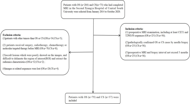
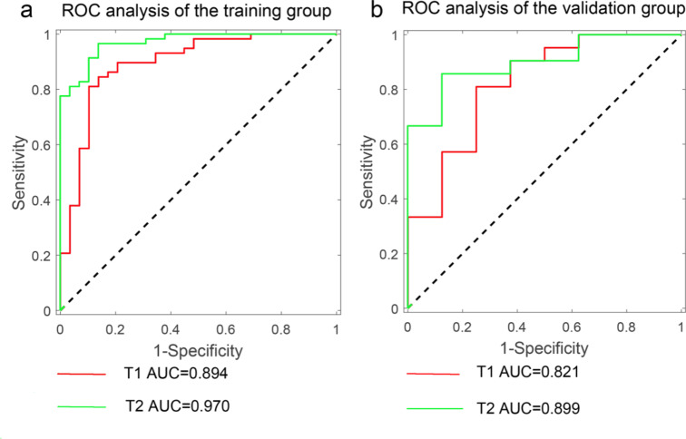
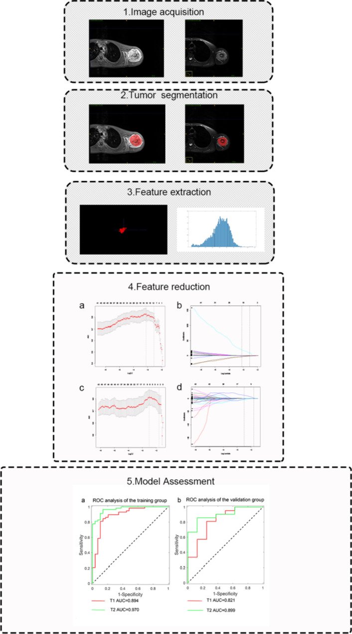
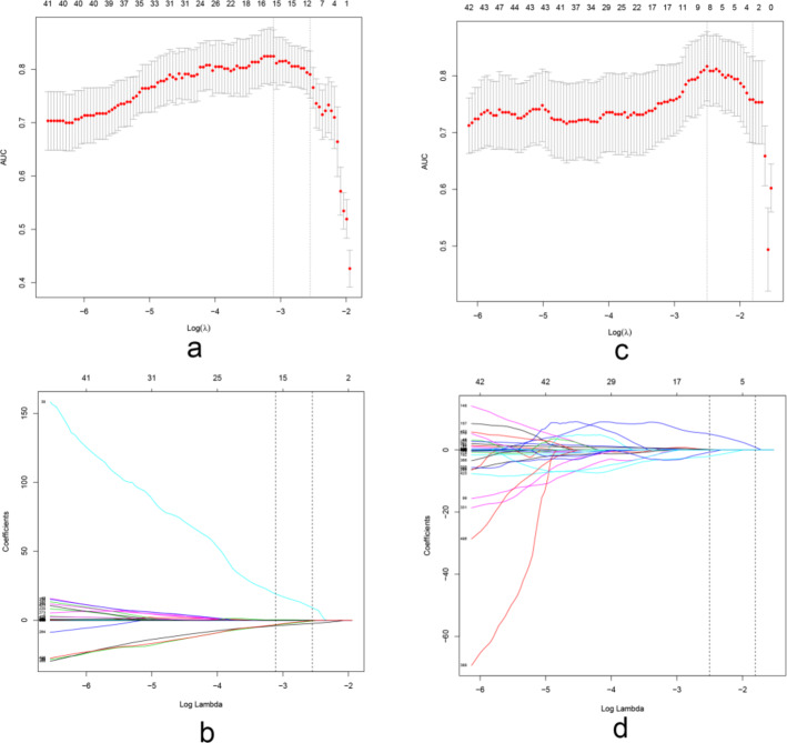
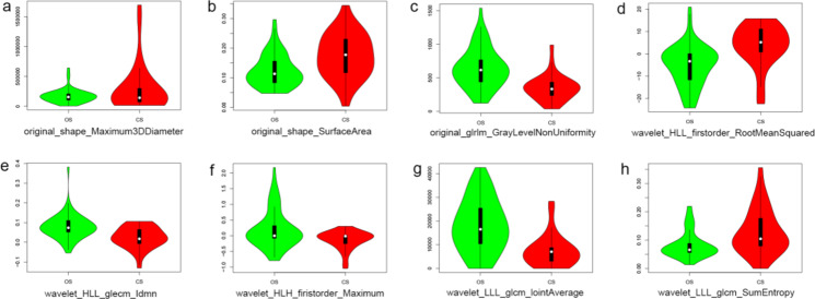
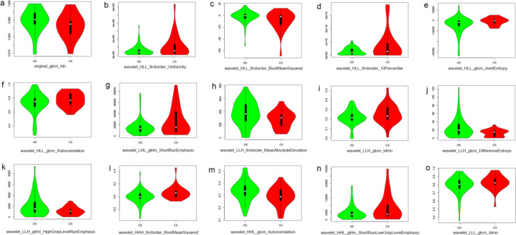
This study was performed to investigate the diagnostic value of radiomics models constructed by fat suppressed T2-weighted imaging (T2WI-FS) and contrast-enhanced T1-weighted imaging (CET1) based on magnetic resonance imaging (MRI) for differentiation of osteosarcoma (OS) and chondrosarcoma (CS). In this retrospective cohort study, we included all inpatients with pathologically confirmed OS or CS from Second Xiangya Hospital of Central South University (Hunan, China) as of October 2020. Demographic and imaging variables were extracted from electronic medical records and compared between OS and CS group. Totals of 530 radiomics features were extracted from CET1 and T2WI-FS sequences based on MRI. The least absolute shrinkage and selection operator (LASSO) method was used for screening and dimensionality reduction of the radiomics model. Multivariate logistic regression analysis was performed to construct the radiomics model, and receiver operating characteristic curve (ROC) was generated to evaluate the diagnostic accuracy of the radiomics model. The training cohort and validation cohort included 87 and 29 patients, respectively. 8 CET1 features and 15 T2WI-FS features were screened based on the radiomics features. In the training group, the area under the receiver-operator characteristic curve (AUC) value for CET1 and T2WI-FS sequences in the radiomics model was 0.894 (95% CI 0.817-0.970) and 0.970 (95% CI 0.940-0.999), respectively. In the validation group, the AUC value for CET1 and T2WI-FS sequences in the radiomics model was 0.821 (95% CI 0.642-1.000) and 0.899 (95% CI 0.785-1.000), respectively. In this study, we developed a radiomics model based on T2WI-FS and CET1 sequences to differentiate between OS and CS. This model exhibits good performance and can help clinicians make decisions and optimize the use of healthcare resources.

摘要

这项研究旨在探讨基于磁共振成像(MRI)的 T2 加权成像(T2WI-FS)和对比增强 T1 加权成像(CET1)构建的放射组学模型在鉴别骨肉瘤(OS)和软骨肉瘤(CS)中的诊断价值。本回顾性队列研究纳入了截至 2020 年 10 月中南大学湘雅二医院(湖南)经病理证实的 OS 或 CS 的所有住院患者。从电子病历中提取人口统计学和影像学变量,并在 OS 和 CS 组之间进行比较。从 CET1 和 T2WI-FS 序列中提取了 530 个放射组学特征。基于 MRI 采用最小绝对值收缩和选择算子(LASSO)方法进行放射组学模型的筛选和降维。采用多变量逻辑回归分析构建放射组学模型,并生成受试者工作特征曲线(ROC)以评估放射组学模型的诊断准确性。训练队列和验证队列分别纳入了 87 例和 29 例患者。基于放射组学特征,筛选出了 8 个 CET1 特征和 15 个 T2WI-FS 特征。在训练组中,该放射组学模型的 CET1 和 T2WI-FS 序列的受试者工作特征曲线(ROC)下面积(AUC)值分别为 0.894(95%CI 0.817-0.970)和 0.970(95%CI 0.940-0.999)。在验证组中,该放射组学模型的 CET1 和 T2WI-FS 序列的 AUC 值分别为 0.821(95%CI 0.642-1.000)和 0.899(95%CI 0.785-1.000)。本研究基于 T2WI-FS 和 CET1 序列构建了一个用于鉴别 OS 和 CS 的放射组学模型。该模型具有良好的性能,可以帮助临床医生做出决策并优化医疗资源的利用。

https://cdn.ncbi.nlm.nih.gov/pmc/blobs/a15b/11535035/05ab34faa884/41598_2024_78245_Fig6_HTML.jpg
https://cdn.ncbi.nlm.nih.gov/pmc/blobs/a15b/11535035/fd8b1fd690d0/41598_2024_78245_Fig1_HTML.jpg
https://cdn.ncbi.nlm.nih.gov/pmc/blobs/a15b/11535035/e7c9e0bc4bac/41598_2024_78245_Fig2_HTML.jpg
https://cdn.ncbi.nlm.nih.gov/pmc/blobs/a15b/11535035/d321759a11a7/41598_2024_78245_Fig3_HTML.jpg
https://cdn.ncbi.nlm.nih.gov/pmc/blobs/a15b/11535035/0ccdf4d710ce/41598_2024_78245_Fig4_HTML.jpg
https://cdn.ncbi.nlm.nih.gov/pmc/blobs/a15b/11535035/232e409761ef/41598_2024_78245_Fig5_HTML.jpg
https://cdn.ncbi.nlm.nih.gov/pmc/blobs/a15b/11535035/05ab34faa884/41598_2024_78245_Fig6_HTML.jpg

相似文献

1
Radiomics analysis in differentiating osteosarcoma and chondrosarcoma based on T2-weighted imaging and contrast-enhanced T1-weighted imaging.基于 T2 加权成像和对比增强 T1 加权成像的影像组学分析在骨肉瘤和软骨肉瘤鉴别诊断中的应用。
Sci Rep. 2024 Nov 4;14(1):26594. doi: 10.1038/s41598-024-78245-1.
2
Differentiation of Pelvic Osteosarcoma and Ewing Sarcoma Using Radiomic Analysis Based on T2-Weighted Images and Contrast-Enhanced T1-Weighted Images.基于 T2 加权图像和对比增强 T1 加权图像的放射组学分析对骨盆骨肉瘤和尤文肉瘤的鉴别诊断。
Biomed Res Int. 2020 May 11;2020:9078603. doi: 10.1155/2020/9078603. eCollection 2020.
3
Preoperative prediction of histopathological grading in patients with chondrosarcoma using MRI-based radiomics with semantic features.基于 MRI 影像组学和语义特征的术前预测软骨肉瘤患者的组织病理学分级。
BMC Med Imaging. 2024 Jul 11;24(1):171. doi: 10.1186/s12880-024-01330-4.
4
Radiomics Nomograms Based on Non-enhanced MRI and Clinical Risk Factors for the Differentiation of Chondrosarcoma from Enchondroma.基于非增强 MRI 及临床危险因素的影像组学列线图模型鉴别软骨肉瘤与内生软骨瘤
J Magn Reson Imaging. 2021 Oct;54(4):1314-1323. doi: 10.1002/jmri.27690. Epub 2021 May 5.
5
[Radiomics models based on non-enhanced MRI can differentiate chondrosarcoma from enchondroma].基于非增强磁共振成像的放射组学模型可鉴别软骨肉瘤与内生软骨瘤
Nan Fang Yi Ke Da Xue Xue Bao. 2020 Apr 30;40(4):483-490. doi: 10.12122/j.issn.1673-4254.2020.04.06.
6
Magnetic resonance imaging based on radiomics for differentiating T1-category nasopharyngeal carcinoma from nasopharyngeal lymphoid hyperplasia: a multicenter study.基于放射组学的磁共振成像鉴别 T1 期鼻咽癌与鼻咽淋巴组织增生:一项多中心研究。
Jpn J Radiol. 2024 Jul;42(7):709-719. doi: 10.1007/s11604-024-01544-0. Epub 2024 Feb 27.
7
Can clinical radiomics nomogram based on 3D multiparametric MRI features and clinical characteristics estimate early recurrence of pelvic chondrosarcoma?基于三维多参数磁共振成像特征和临床特征的临床影像组学列线图能否评估骨盆软骨肉瘤的早期复发情况?
J Magn Reson Imaging. 2020 Feb;51(2):435-445. doi: 10.1002/jmri.26834. Epub 2019 Jun 18.
8
Development and external validation of an MRI-based radiomics nomogram for pretreatment prediction for early relapse in osteosarcoma: A retrospective multicenter study.基于 MRI 的放射组学列线图预测骨肉瘤早期复发的建立与外部验证:一项回顾性多中心研究。
Eur J Radiol. 2020 Aug;129:109066. doi: 10.1016/j.ejrad.2020.109066. Epub 2020 May 17.
9
Pretreatment MR imaging radiomics signatures for response prediction to induction chemotherapy in patients with nasopharyngeal carcinoma.鼻咽癌患者诱导化疗反应预测的预处理 MRI 影像组学特征。
Eur J Radiol. 2018 Jan;98:100-106. doi: 10.1016/j.ejrad.2017.11.007. Epub 2017 Nov 14.
10
A Triple-Classification Radiomics Model for the Differentiation of Primary Chordoma, Giant Cell Tumor, and Metastatic Tumor of Sacrum Based on T2-Weighted and Contrast-Enhanced T1-Weighted MRI.基于 T2 加权和增强 T1 加权 MRI 的原发性脊索瘤、巨细胞瘤和骶骨转移瘤的三重分类放射组学模型。
J Magn Reson Imaging. 2019 Mar;49(3):752-759. doi: 10.1002/jmri.26238. Epub 2018 Nov 14.

引用本文的文献

1
Multidimensional bioinformatics analysis of chondrosarcoma subtypes and TGF-β signaling networks using big data approaches.使用大数据方法对软骨肉瘤亚型和转化生长因子-β信号网络进行多维度生物信息学分析。
Discov Oncol. 2025 Jun 17;16(1):1136. doi: 10.1007/s12672-025-02931-3.
2
Building a Therapeutic Bridge Between Dogs and Humans: A Review of Potential Cross-Species Osteosarcoma Biomarkers.搭建犬类与人类之间的治疗桥梁:潜在跨物种骨肉瘤生物标志物综述
Int J Mol Sci. 2025 May 28;26(11):5152. doi: 10.3390/ijms26115152.
3
Interpretable machine learning and radiomics in hip MRI diagnostics: comparing ONFH and OA predictions to experts.

本文引用的文献

1
Effect of Cisplatin Arterial Infusion (CAI) on Primary Nonmetastatic Pelvic Osteosarcoma: A Preliminary Study.顺铂动脉灌注(CAI)对原发性非转移性骨盆骨肉瘤的影响:一项初步研究。
Cancer Manag Res. 2021 Feb 15;13:1491-1503. doi: 10.2147/CMAR.S294677. eCollection 2021.
2
Noncoding RNAs in osteosarcoma: Implications for drug resistance.非编码 RNA 在骨肉瘤中的作用:与耐药性的关系。
Cancer Lett. 2021 Apr 28;504:91-103. doi: 10.1016/j.canlet.2021.02.007. Epub 2021 Feb 12.
3
MDCT-Based Radiomics Features for the Differentiation of Serous Borderline Ovarian Tumors and Serous Malignant Ovarian Tumors.
髋关节MRI诊断中的可解释机器学习与放射组学:将股骨头缺血性坏死和骨关节炎预测结果与专家进行比较。
Front Immunol. 2025 Jan 29;16:1532248. doi: 10.3389/fimmu.2025.1532248. eCollection 2025.
基于多层螺旋CT的影像组学特征用于鉴别浆液性交界性卵巢肿瘤和浆液性恶性卵巢肿瘤
Cancer Manag Res. 2021 Jan 12;13:329-336. doi: 10.2147/CMAR.S284220. eCollection 2021.
4
Preoperative classification of primary and metastatic liver cancer via machine learning-based ultrasound radiomics.基于机器学习的超声放射组学对原发性和转移性肝癌的术前分类。
Eur Radiol. 2021 Jul;31(7):4576-4586. doi: 10.1007/s00330-020-07562-6. Epub 2021 Jan 14.
5
Radiomic signature based on CT imaging to distinguish invasive adenocarcinoma from minimally invasive adenocarcinoma in pure ground-glass nodules with pleural contact.基于 CT 成像的放射组学特征区分胸膜接触的纯磨玻璃结节中浸润性腺癌与微浸润性腺癌
Cancer Imaging. 2021 Jan 6;21(1):1. doi: 10.1186/s40644-020-00376-1.
6
Distinguishing brain inflammation from grade II glioma in population without contrast enhancement: a radiomics analysis based on conventional MRI.在无对比增强的人群中区分脑炎症与 II 级胶质瘤:基于常规 MRI 的放射组学分析。
Eur J Radiol. 2021 Jan;134:109467. doi: 10.1016/j.ejrad.2020.109467. Epub 2020 Dec 3.
7
Can computed tomography-based radiomics potentially discriminate between anterior mediastinal cysts and type B1 and B2 thymomas?基于计算机断层扫描的放射组学是否有可能区分前纵隔囊肿与 B1 型和 B2 型胸腺瘤?
Biomed Eng Online. 2020 Nov 27;19(1):89. doi: 10.1186/s12938-020-00833-9.
8
Chondroblastic osteosarcoma-A case report and review of literature.软骨母细胞性骨肉瘤——病例报告及文献综述
Clin Case Rep. 2019 Sep 30;8(11):2097-2102. doi: 10.1002/ccr3.1761. eCollection 2020 Nov.
9
MRI-Based radiomics nomogram for differentiation of benign and malignant lesions of the parotid gland.基于 MRI 的影像组学列线图用于鉴别腮腺良恶性病变。
Eur Radiol. 2021 Jun;31(6):4042-4052. doi: 10.1007/s00330-020-07483-4. Epub 2020 Nov 19.
10
Prediction of the clinicopathological subtypes of breast cancer using a fisher discriminant analysis model based on radiomic features of diffusion-weighted MRI.基于扩散加权 MRI 影像组学特征的 Fisher 判别分析模型预测乳腺癌的临床病理亚型。
BMC Cancer. 2020 Nov 9;20(1):1073. doi: 10.1186/s12885-020-07557-y.