• 文献检索
  • 文档翻译
  • 深度研究
  • 学术资讯
  • Suppr Zotero 插件Zotero 插件
  • 邀请有礼
  • 套餐&价格
  • 历史记录
应用&插件
Suppr Zotero 插件Zotero 插件浏览器插件Mac 客户端Windows 客户端微信小程序
定价
高级版会员购买积分包购买API积分包
服务
文献检索文档翻译深度研究API 文档MCP 服务
关于我们
关于 Suppr公司介绍联系我们用户协议隐私条款
关注我们

Suppr 超能文献

核心技术专利:CN118964589B侵权必究
粤ICP备2023148730 号-1Suppr @ 2026

文献检索

告别复杂PubMed语法,用中文像聊天一样搜索,搜遍4000万医学文献。AI智能推荐,让科研检索更轻松。

立即免费搜索

文件翻译

保留排版,准确专业,支持PDF/Word/PPT等文件格式,支持 12+语言互译。

免费翻译文档

深度研究

AI帮你快速写综述,25分钟生成高质量综述,智能提取关键信息,辅助科研写作。

立即免费体验

基于扩散加权 MRI 影像组学特征的 Fisher 判别分析模型预测乳腺癌的临床病理亚型。

Prediction of the clinicopathological subtypes of breast cancer using a fisher discriminant analysis model based on radiomic features of diffusion-weighted MRI.

机构信息

Department of Radiology, The Affiliated Hospital of Qingdao University, No.59 Haier Road, Qingdao, 266000, China.

Department of Pediatric Surgery, Shandong University Qilu Hospital, Jinan, 250012, China.

出版信息

BMC Cancer. 2020 Nov 9;20(1):1073. doi: 10.1186/s12885-020-07557-y.

DOI:10.1186/s12885-020-07557-y
PMID:33167903
原文链接:https://pmc.ncbi.nlm.nih.gov/articles/PMC7654148/
Abstract

BACKGROUND

The clinicopathological classification of breast cancer is proposed according to therapeutic purposes. It is simplified and can be conducted easily in clinical practice, and this subtyping undoubtedly contributes to the treatment selection of breast cancer. This study aims to investigate the feasibility of using a Fisher discriminant analysis model based on radiomic features of diffusion-weighted MRI for predicting the clinicopathological subtypes of breast cancer.

METHODS

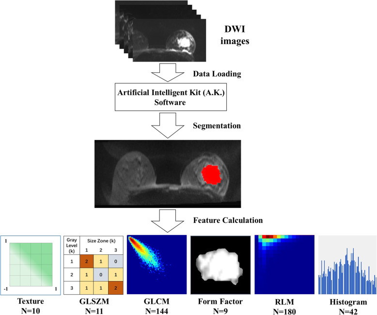
Patients who underwent breast magnetic resonance imaging were confirmed by retrieving data from our institutional picture archiving and communication system (PACS) between March 2013 and September 2017. Five clinicopathological subtypes were determined based on the status of ER, PR, HER2 and Ki-67 from the immunohistochemical test. The radiomic features of diffusion-weighted imaging were derived from the volume of interest (VOI) of each tumour. Fisher discriminant analysis was performed for clinicopathological subtyping by using a backward selection method. To evaluate the diagnostic performance of the radiomic features, ROC analyses were performed to differentiate between immunohistochemical biomarker-positive and -negative groups.

RESULTS

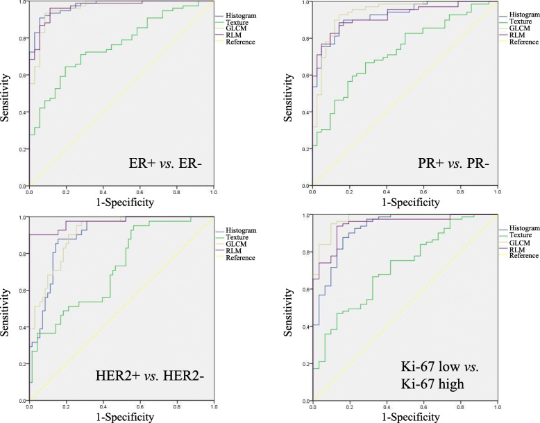
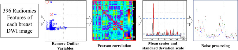
A total of 84 radiomic features of four statistical methods were included after preprocessing. The overall accuracy for predicting the clinicopathological subtypes was 96.4% by Fisher discriminant analysis, and the weighted accuracy was 96.6%. For predicting diverse clinicopathological subtypes, the prediction accuracies ranged from 92 to 100%. According to the cross-validation, the overall accuracy of the model was 82.1%, and the accuracies of the model for predicting the luminal A, luminal B, luminal B, HER2 positive and triple negative subtypes were 79, 77, 88, 92 and 73%, respectively. According to the ROC analysis, the radiomic features had excellent performance in differentiating between different statuses of ER, PR, HER2 and Ki-67.

CONCLUSIONS

The Fisher discriminant analysis model based on radiomic features of diffusion-weighted MRI is a reliable method for the prediction of clinicopathological breast cancer subtypes.

摘要

背景

乳腺癌的临床病理分类是根据治疗目的提出的。它简化了临床实践中的操作,这种分型无疑有助于乳腺癌的治疗选择。本研究旨在探讨基于扩散加权 MRI 影像组学特征的 Fisher 判别分析模型预测乳腺癌临床病理亚型的可行性。

方法

通过检索我院影像归档和通信系统(PACS)2013 年 3 月至 2017 年 9 月的数据,确定接受乳腺磁共振成像检查的患者。根据免疫组织化学检查中 ER、PR、HER2 和 Ki-67 的状态确定五种临床病理亚型。从每个肿瘤的感兴趣区域(VOI)中提取扩散加权成像的影像组学特征。采用后向选择法进行 Fisher 判别分析以进行临床病理分型。为了评估影像组学特征的诊断性能,通过 ROC 分析对免疫组化生物标志物阳性和阴性组进行了区分。

结果

预处理后共纳入 84 个来自四种统计方法的影像组学特征。Fisher 判别分析预测临床病理亚型的总准确率为 96.4%,加权准确率为 96.6%。对于预测不同的临床病理亚型,预测准确率范围为 92%至 100%。根据交叉验证,该模型的总准确率为 82.1%,预测 luminal A、luminal B、luminal B、HER2 阳性和三阴性亚型的准确率分别为 79%、77%、88%、92%和 73%。根据 ROC 分析,影像组学特征在区分 ER、PR、HER2 和 Ki-67 的不同状态方面具有优异的性能。

结论

基于扩散加权 MRI 影像组学特征的 Fisher 判别分析模型是预测乳腺癌临床病理亚型的可靠方法。

https://cdn.ncbi.nlm.nih.gov/pmc/blobs/20de/7654148/b20f951a1b3b/12885_2020_7557_Fig3_HTML.jpg
https://cdn.ncbi.nlm.nih.gov/pmc/blobs/20de/7654148/302302cd8526/12885_2020_7557_Fig1_HTML.jpg
https://cdn.ncbi.nlm.nih.gov/pmc/blobs/20de/7654148/39817627a707/12885_2020_7557_Fig2_HTML.jpg
https://cdn.ncbi.nlm.nih.gov/pmc/blobs/20de/7654148/b20f951a1b3b/12885_2020_7557_Fig3_HTML.jpg
https://cdn.ncbi.nlm.nih.gov/pmc/blobs/20de/7654148/302302cd8526/12885_2020_7557_Fig1_HTML.jpg
https://cdn.ncbi.nlm.nih.gov/pmc/blobs/20de/7654148/39817627a707/12885_2020_7557_Fig2_HTML.jpg
https://cdn.ncbi.nlm.nih.gov/pmc/blobs/20de/7654148/b20f951a1b3b/12885_2020_7557_Fig3_HTML.jpg

相似文献

1
Prediction of the clinicopathological subtypes of breast cancer using a fisher discriminant analysis model based on radiomic features of diffusion-weighted MRI.基于扩散加权 MRI 影像组学特征的 Fisher 判别分析模型预测乳腺癌的临床病理亚型。
BMC Cancer. 2020 Nov 9;20(1):1073. doi: 10.1186/s12885-020-07557-y.
2
Radiomic signatures with contrast-enhanced magnetic resonance imaging for the assessment of breast cancer receptor status and molecular subtypes: initial results.基于对比增强磁共振成像的放射组学特征用于评估乳腺癌受体状态和分子亚型:初步结果。
Breast Cancer Res. 2019 Sep 12;21(1):106. doi: 10.1186/s13058-019-1187-z.
3
Radiomic Signatures Derived from Diffusion-Weighted Imaging for the Assessment of Breast Cancer Receptor Status and Molecular Subtypes.基于扩散加权成像的影像组学特征评估乳腺癌受体状态和分子亚型。
Mol Imaging Biol. 2020 Apr;22(2):453-461. doi: 10.1007/s11307-019-01383-w.
4
Diffusion-weighted imaging (DWI) with apparent diffusion coefficient (ADC) mapping as a quantitative imaging biomarker for prediction of immunohistochemical receptor status, proliferation rate, and molecular subtypes of breast cancer.弥散加权成像(DWI)及其表观扩散系数(ADC)图作为一种定量影像学生物标志物,可预测乳腺癌的免疫组织化学受体状态、增殖率和分子亚型。
J Magn Reson Imaging. 2019 Sep;50(3):836-846. doi: 10.1002/jmri.26697. Epub 2019 Feb 27.
5
Radiomic analysis on magnetic resonance diffusion weighted image in distinguishing triple-negative breast cancer from other subtypes: a feasibility study.磁共振弥散加权图像纹理分析鉴别三阴性乳腺癌与其他亚型的可行性研究。
Clin Imaging. 2021 Apr;72:136-141. doi: 10.1016/j.clinimag.2020.11.024. Epub 2020 Nov 14.
6
Breast MRI background parenchymal enhancement as an imaging bridge to molecular cancer sub-type.乳腺 MRI 背景实质增强作为连接分子肿瘤亚型的影像学桥梁。
Eur J Radiol. 2019 Apr;113:148-152. doi: 10.1016/j.ejrad.2019.02.018. Epub 2019 Feb 15.
7
Long-Term Outcomes of Immunohistochemically Defined Subtypes of Breast Cancer Less Than or Equal to 2 cm After Breast-Conserving Surgery.保乳手术后最大直径小于或等于 2cm 的乳腺癌免疫组化定义亚型的长期预后
J Surg Res. 2019 Apr;236:288-299. doi: 10.1016/j.jss.2018.11.028. Epub 2018 Dec 27.
8
Clinicopathological features of breast cancer with different molecular subtypes in Chinese women.中国女性不同分子亚型乳腺癌的临床病理特征
J Huazhong Univ Sci Technolog Med Sci. 2013 Feb;33(1):117-121. doi: 10.1007/s11596-013-1082-2. Epub 2013 Feb 8.
9
[Clinical characteristics and survival in the operable breast cancer patients with different molecular subtypes].[不同分子亚型可手术乳腺癌患者的临床特征与生存情况]
Zhonghua Zhong Liu Za Zhi. 2009 Jun;31(6):447-51.
10
Breast Cancer Outcomes as Defined by the Estrogen Receptor, Progesterone Receptor, and Human Growth Factor Receptor-2 in a Multi-ethnic Asian Country.在一个多民族亚洲国家中,由雌激素受体、孕激素受体和人类生长因子受体-2所定义的乳腺癌预后情况。
World J Surg. 2015 Oct;39(10):2450-8. doi: 10.1007/s00268-015-3133-2.

引用本文的文献

1
Performance of low- and high-temporal-resolution DCE-MRI texture analysis in distinguishing breast lesions from background enhancement.低时间分辨率和高时间分辨率动态对比增强磁共振成像(DCE-MRI)纹理分析在区分乳腺病变与背景强化方面的性能
Am J Transl Res. 2025 Aug 15;17(8):6676-6687. doi: 10.62347/KKUZ9662. eCollection 2025.
2
Radiologic imaging biomarkers in triple-negative breast cancer: a literature review about the role of artificial intelligence and the way forward.三阴性乳腺癌的放射影像学生物标志物:关于人工智能作用及未来发展方向的文献综述
BJR Artif Intell. 2024 Nov 13;1(1):ubae016. doi: 10.1093/bjrai/ubae016. eCollection 2024 Jan.
3

本文引用的文献

1
Non-Invasive Assessment of Breast Cancer Molecular Subtypes with Multiparametric Magnetic Resonance Imaging Radiomics.基于多参数磁共振成像放射组学的乳腺癌分子亚型无创评估
J Clin Med. 2020 Jun 14;9(6):1853. doi: 10.3390/jcm9061853.
2
Diffusion-weighted imaging or dynamic contrast-enhanced curve: a retrospective analysis of contrast-enhanced magnetic resonance imaging-based differential diagnoses of benign and malignant breast lesions.弥散加权成像或动态对比增强曲线:基于对比增强磁共振成像的良性和恶性乳腺病变鉴别诊断的回顾性分析。
Eur Radiol. 2020 Sep;30(9):4795-4805. doi: 10.1007/s00330-020-06883-w. Epub 2020 Apr 29.
3
Radiomics analysis in differentiating osteosarcoma and chondrosarcoma based on T2-weighted imaging and contrast-enhanced T1-weighted imaging.
基于 T2 加权成像和对比增强 T1 加权成像的影像组学分析在骨肉瘤和软骨肉瘤鉴别诊断中的应用。
Sci Rep. 2024 Nov 4;14(1):26594. doi: 10.1038/s41598-024-78245-1.
4
Predicting HER2 Status Associated with Breast Cancer Aggressiveness Using Four Machine Learning Models.使用四种机器学习模型预测与乳腺癌侵袭性相关的 HER2 状态。
Asian Pac J Cancer Prev. 2024 Oct 1;25(10):3609-3618. doi: 10.31557/APJCP.2024.25.10.3609.
5
Differences in Histological Subtypes of Invasive Lobular Breast Carcinoma According to Immunohistochemical Molecular Classification.根据免疫组织化学分子分类的浸润性小叶乳腺癌组织学亚型差异
Diagnostics (Basel). 2024 Mar 21;14(6):660. doi: 10.3390/diagnostics14060660.
6
The use of radiomics in magnetic resonance imaging for the pre-treatment characterisation of breast cancers: A scoping review.磁共振成像中放射组学在乳腺癌治疗前特征描述中的应用:范围综述。
J Med Radiat Sci. 2023 Dec;70(4):462-478. doi: 10.1002/jmrs.709. Epub 2023 Aug 3.
7
Histopathological Images Analysis and Predictive Modeling Implemented in Digital Pathology-Current Affairs and Perspectives.数字病理学中实施的组织病理学图像分析与预测建模——现状与展望
Diagnostics (Basel). 2023 Jul 14;13(14):2379. doi: 10.3390/diagnostics13142379.
8
A Two-Step Feature Selection Radiomic Approach to Predict Molecular Outcomes in Breast Cancer.两步式特征选择放射组学方法预测乳腺癌分子预后。
Sensors (Basel). 2023 Jan 31;23(3):1552. doi: 10.3390/s23031552.
9
Fisher discriminant model based on LASSO logistic regression for computed tomography imaging diagnosis of pelvic rhabdomyosarcoma in children.基于 LASSO 逻辑回归的 Fisher 判别模型在儿童骨盆横纹肌肉瘤 CT 影像诊断中的应用。
Sci Rep. 2022 Sep 17;12(1):15631. doi: 10.1038/s41598-022-20051-8.
10
Prediction of carotid plaque by blood biochemical indices and related factors based on Fisher discriminant analysis.基于 Fisher 判别分析的血生化指标及相关因素对颈动脉斑块的预测。
BMC Cardiovasc Disord. 2022 Aug 15;22(1):371. doi: 10.1186/s12872-022-02806-3.
Diffusion weighted magnetic resonance imaging (DW-MRI) as a non-invasive, tissue cellularity marker to monitor cancer treatment response.
弥散加权磁共振成像(DW-MRI)作为一种非侵入性的组织细胞标志物,可用于监测癌症治疗反应。
BMC Cancer. 2020 Feb 19;20(1):134. doi: 10.1186/s12885-020-6617-x.
4
Breast Cancer Radiogenomics: Current Status and Future Directions.乳腺癌放射组学:现状与未来方向。
Acad Radiol. 2020 Jan;27(1):39-46. doi: 10.1016/j.acra.2019.09.012.
5
Approach for the Definition of radiomiRNomic Signatures for Breast Cancer Differential Diagnosis.用于乳腺癌鉴别诊断的 radiomiRNomic 特征定义方法。
Int J Mol Sci. 2019 Nov 20;20(23):5825. doi: 10.3390/ijms20235825.
6
Tumour heterogeneity revealed by unsupervised decomposition of dynamic contrast-enhanced magnetic resonance imaging is associated with underlying gene expression patterns and poor survival in breast cancer patients.非监督性动态对比增强磁共振成像分解所揭示的肿瘤异质性与乳腺癌患者的潜在基因表达模式和不良预后相关。
Breast Cancer Res. 2019 Oct 17;21(1):112. doi: 10.1186/s13058-019-1199-8.
7
Diffusion-weighted MRI for Unenhanced Breast Cancer Screening.弥散加权 MRI 在乳腺癌筛查中的应用。
Radiology. 2019 Dec;293(3):504-520. doi: 10.1148/radiol.2019182789. Epub 2019 Oct 8.
8
Radiogenomics of breast cancer using dynamic contrast enhanced MRI and gene expression profiling.基于动态对比增强 MRI 和基因表达谱的乳腺癌放射组学研究。
Cancer Imaging. 2019 Jul 15;19(1):48. doi: 10.1186/s40644-019-0233-5.
9
Machine Learning-Based Analysis of MR Multiparametric Radiomics for the Subtype Classification of Breast Cancer.基于机器学习的磁共振多参数放射组学对乳腺癌亚型分类的分析
Front Oncol. 2019 Jun 14;9:505. doi: 10.3389/fonc.2019.00505. eCollection 2019.
10
Radiomic Signatures Derived from Diffusion-Weighted Imaging for the Assessment of Breast Cancer Receptor Status and Molecular Subtypes.基于扩散加权成像的影像组学特征评估乳腺癌受体状态和分子亚型。
Mol Imaging Biol. 2020 Apr;22(2):453-461. doi: 10.1007/s11307-019-01383-w.