• 文献检索
  • 文档翻译
  • 深度研究
  • 学术资讯
  • Suppr Zotero 插件Zotero 插件
  • 邀请有礼
  • 套餐&价格
  • 历史记录
应用&插件
Suppr Zotero 插件Zotero 插件浏览器插件Mac 客户端Windows 客户端微信小程序
定价
高级版会员购买积分包购买API积分包
服务
文献检索文档翻译深度研究API 文档MCP 服务
关于我们
关于 Suppr公司介绍联系我们用户协议隐私条款
关注我们

Suppr 超能文献

核心技术专利:CN118964589B侵权必究
粤ICP备2023148730 号-1Suppr @ 2026

文献检索

告别复杂PubMed语法,用中文像聊天一样搜索,搜遍4000万医学文献。AI智能推荐,让科研检索更轻松。

立即免费搜索

文件翻译

保留排版,准确专业,支持PDF/Word/PPT等文件格式,支持 12+语言互译。

免费翻译文档

深度研究

AI帮你快速写综述,25分钟生成高质量综述,智能提取关键信息,辅助科研写作。

立即免费体验

基于 T2 加权图像和对比增强 T1 加权图像的放射组学分析对骨盆骨肉瘤和尤文肉瘤的鉴别诊断。

Differentiation of Pelvic Osteosarcoma and Ewing Sarcoma Using Radiomic Analysis Based on T2-Weighted Images and Contrast-Enhanced T1-Weighted Images.

机构信息

Department of Radiology, Peking University People's Hospital, Beijing, China 100044.

Department of Radiology, Peking University Shenzhen Hospital, Shenzhen, Guangdong, China 518036.

出版信息

Biomed Res Int. 2020 May 11;2020:9078603. doi: 10.1155/2020/9078603. eCollection 2020.

DOI:10.1155/2020/9078603
PMID:32462033
原文链接:https://pmc.ncbi.nlm.nih.gov/articles/PMC7240794/
Abstract

OBJECTIVE

To determine if osteosarcoma (OS) and Ewing sarcoma (EWS) of the pelvis based on MRI can be differentiated using radiomic analysis.

MATERIALS AND METHODS

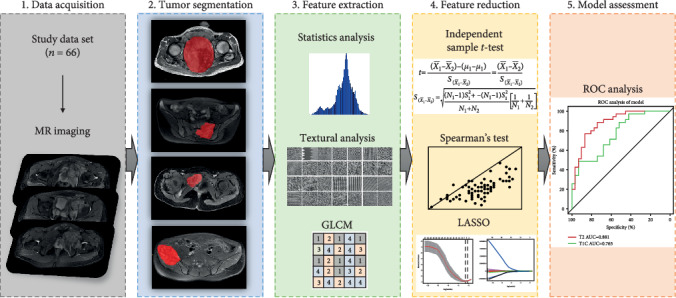
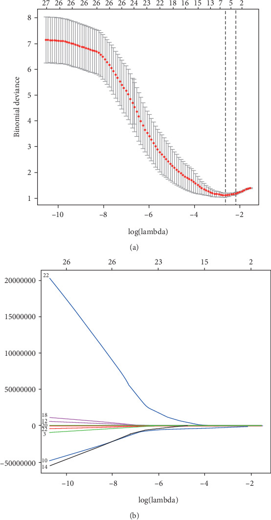
In this study, 3.0 T magnetic resonance (MR) data of 66 patients (40 males and 26 females, mean age 27.6 ± 13.9 years) with pathologically confirmed OS or EWS of the pelvis (35 with OS and 31 with EWS) taken from April 2013 to December 2017 were retrospectively reviewed. T2-weighted fat-saturated (T2-FS) and contrast-enhanced T1-weighted (CET1) images were manually segmented, and imaging features were extracted. Independent-sample -test, Spearman's test, and the least absolute shrinkage and selection operator (LASSO) method were used to select the most useful features from the original data set. The performance of radiomic analysis was investigated by the area under the receiver operating characteristic (ROC) curve (AUC) analysis.

RESULTS

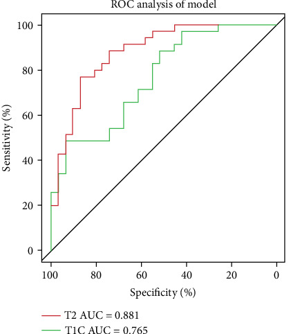
385 initial features were extracted from T2-FS and CET1 MR data. Nine features from T2-FS and 7 features from CET1 were selected by using the LASSO method. The radiomic analysis to differentiate OS and EWS of the pelvis based on T2-FS and CET1 images using the aforementioned selected features achieved AUC values of 0.881 (95% confidence interval (CI): 0.799-0.963) and 0.765 (95% CI: 0.652-0.878), respectively.

CONCLUSION

Radiomic analysis showed potential in differentiating OS from EWS of the pelvis, in which T2-FS demonstrated better diagnostic value. To differentiate OS from EWS of the pelvis using our multiparametric MRI-based radiomic analysis could preoperatively improve diagnostic accuracy and greatly contribute to therapy planning.

摘要

目的

通过放射组学分析,确定骨盆骨肉瘤(OS)和尤因肉瘤(EWS)是否可以基于 MRI 进行区分。

材料和方法

本研究回顾性分析了 2013 年 4 月至 2017 年 12 月期间,经病理证实的骨盆 OS 或 EWS 的 66 例患者(男性 40 例,女性 26 例,平均年龄 27.6±13.9 岁)的 3.0T 磁共振(MR)数据(35 例为 OS,31 例为 EWS)。对 T2 加权脂肪饱和(T2-FS)和对比增强 T1 加权(CET1)图像进行手动分割,并提取成像特征。采用独立样本 t 检验、Spearman 检验和最小绝对收缩和选择算子(LASSO)方法,从原始数据集选择最有用的特征。通过受试者工作特征(ROC)曲线(AUC)分析评估放射组学分析的性能。

结果

从 T2-FS 和 CET1MR 数据中提取了 385 个初始特征。通过 LASSO 方法,从 T2-FS 中选择了 9 个特征,从 CET1 中选择了 7 个特征。基于上述选定特征,利用 T2-FS 和 CET1 图像对骨盆 OS 和 EWS 进行鉴别分析的放射组学分析,AUC 值分别为 0.881(95%置信区间(CI):0.799-0.963)和 0.765(95%CI:0.652-0.878)。

结论

放射组学分析显示出区分骨盆 OS 和 EWS 的潜力,其中 T2-FS 显示出更好的诊断价值。利用我们的基于多参数 MRI 的放射组学分析对骨盆 OS 和 EWS 进行鉴别,可在术前提高诊断准确性,并极大地有助于治疗计划。

https://cdn.ncbi.nlm.nih.gov/pmc/blobs/cc86/7240794/fd6abc3f9b5b/BMRI2020-9078603.003.jpg
https://cdn.ncbi.nlm.nih.gov/pmc/blobs/cc86/7240794/2f7ef7c32b25/BMRI2020-9078603.001.jpg
https://cdn.ncbi.nlm.nih.gov/pmc/blobs/cc86/7240794/7e9696b32b0a/BMRI2020-9078603.002.jpg
https://cdn.ncbi.nlm.nih.gov/pmc/blobs/cc86/7240794/fd6abc3f9b5b/BMRI2020-9078603.003.jpg
https://cdn.ncbi.nlm.nih.gov/pmc/blobs/cc86/7240794/2f7ef7c32b25/BMRI2020-9078603.001.jpg
https://cdn.ncbi.nlm.nih.gov/pmc/blobs/cc86/7240794/7e9696b32b0a/BMRI2020-9078603.002.jpg
https://cdn.ncbi.nlm.nih.gov/pmc/blobs/cc86/7240794/fd6abc3f9b5b/BMRI2020-9078603.003.jpg

相似文献

1
Differentiation of Pelvic Osteosarcoma and Ewing Sarcoma Using Radiomic Analysis Based on T2-Weighted Images and Contrast-Enhanced T1-Weighted Images.基于 T2 加权图像和对比增强 T1 加权图像的放射组学分析对骨盆骨肉瘤和尤文肉瘤的鉴别诊断。
Biomed Res Int. 2020 May 11;2020:9078603. doi: 10.1155/2020/9078603. eCollection 2020.
2
Development and external validation of an MRI-based radiomics nomogram for pretreatment prediction for early relapse in osteosarcoma: A retrospective multicenter study.基于 MRI 的放射组学列线图预测骨肉瘤早期复发的建立与外部验证:一项回顾性多中心研究。
Eur J Radiol. 2020 Aug;129:109066. doi: 10.1016/j.ejrad.2020.109066. Epub 2020 May 17.
3
Prediction of malignant glioma grades using contrast-enhanced T1-weighted and T2-weighted magnetic resonance images based on a radiomic analysis.基于放射组学分析的对比增强 T1 加权和 T2 加权磁共振成像预测恶性胶质瘤分级。
Sci Rep. 2019 Dec 19;9(1):19411. doi: 10.1038/s41598-019-55922-0.
4
Radiomics-Based Pretherapeutic Prediction of Non-response to Neoadjuvant Therapy in Locally Advanced Rectal Cancer.基于放射组学的局部晚期直肠癌新辅助治疗无反应的预测。
Ann Surg Oncol. 2019 Jun;26(6):1676-1684. doi: 10.1245/s10434-019-07300-3. Epub 2019 Mar 18.
5
A Triple-Classification Radiomics Model for the Differentiation of Primary Chordoma, Giant Cell Tumor, and Metastatic Tumor of Sacrum Based on T2-Weighted and Contrast-Enhanced T1-Weighted MRI.基于 T2 加权和增强 T1 加权 MRI 的原发性脊索瘤、巨细胞瘤和骶骨转移瘤的三重分类放射组学模型。
J Magn Reson Imaging. 2019 Mar;49(3):752-759. doi: 10.1002/jmri.26238. Epub 2018 Nov 14.
6
Differentiating axillary lymph node metastasis in invasive breast cancer patients: A comparison of radiomic signatures from multiparametric breast MR sequences.鉴别浸润性乳腺癌患者腋窝淋巴结转移:多参数乳腺磁共振序列的放射组学特征比较。
J Magn Reson Imaging. 2019 Oct;50(4):1125-1132. doi: 10.1002/jmri.26701. Epub 2019 Mar 7.
7
The diagnostic value of magnetic resonance imaging-based texture analysis in differentiating enchondroma and chondrosarcoma.基于磁共振成像的纹理分析在鉴别内生软骨瘤和软骨肉瘤中的诊断价值。
Skeletal Radiol. 2023 May;52(5):1039-1049. doi: 10.1007/s00256-022-04242-y. Epub 2022 Nov 25.
8
Machine Learning-Based Multiparametric Magnetic Resonance Imaging Radiomic Model for Discrimination of Pathological Subtypes of Craniopharyngioma.基于机器学习的多参数磁共振成像放射组学模型用于鉴别颅咽管瘤的病理亚型
J Magn Reson Imaging. 2021 Nov;54(5):1541-1550. doi: 10.1002/jmri.27761. Epub 2021 Jun 4.
9
Radiomic nomogram based on MRI to predict grade of branching type intraductal papillary mucinous neoplasms of the pancreas: a multicenter study.基于 MRI 的放射组学列线图预测胰腺分支型管状乳头状黏液性肿瘤的分级:一项多中心研究。
Cancer Imaging. 2021 Mar 9;21(1):26. doi: 10.1186/s40644-021-00395-6.
10
Percent slope analysis of dynamic magnetic resonance imaging for assessment of chemotherapy response of osteosarcoma or Ewing sarcoma: systematic review and meta-analysis.动态磁共振成像的百分比斜率分析用于评估骨肉瘤或尤因肉瘤的化疗反应:系统评价与荟萃分析
Skeletal Radiol. 2016 Sep;45(9):1235-42. doi: 10.1007/s00256-016-2410-y. Epub 2016 May 26.

引用本文的文献

1
Integrating radiomics, artificial intelligence, and molecular signatures in bone and soft tissue tumors: advances in diagnosis and prognostication.整合放射组学、人工智能和分子特征于骨与软组织肿瘤:诊断与预后的进展
Front Oncol. 2025 Aug 18;15:1613133. doi: 10.3389/fonc.2025.1613133. eCollection 2025.
2
Radiomics analysis in differentiating osteosarcoma and chondrosarcoma based on T2-weighted imaging and contrast-enhanced T1-weighted imaging.基于 T2 加权成像和对比增强 T1 加权成像的影像组学分析在骨肉瘤和软骨肉瘤鉴别诊断中的应用。
Sci Rep. 2024 Nov 4;14(1):26594. doi: 10.1038/s41598-024-78245-1.
3
Preoperative prediction of high-grade osteosarcoma response to neoadjuvant therapy based on a plain CT radiomics model: A dual-center study.

本文引用的文献

1
Intravoxel incoherent motion diffusion-weighted MR imaging for differentiation of benign and malignant musculoskeletal tumours at 3 T.3T磁共振体素内不相干运动扩散加权成像用于鉴别骨骼肌肉系统良恶性肿瘤
Br J Radiol. 2018 Feb;91(1082):20170636. doi: 10.1259/bjr.20170636. Epub 2017 Dec 15.
2
Tumor radiomic heterogeneity: Multiparametric functional imaging to characterize variability and predict response following cervical cancer radiation therapy.肿瘤放射组学异质性:多参数功能成像以描述宫颈癌放疗后变异性和预测反应。
J Magn Reson Imaging. 2018 May;47(5):1388-1396. doi: 10.1002/jmri.25874. Epub 2017 Oct 16.
3
基于平扫CT影像组学模型的高级别骨肉瘤对新辅助治疗反应的术前预测:一项双中心研究
J Bone Oncol. 2024 Jun 8;47:100614. doi: 10.1016/j.jbo.2024.100614. eCollection 2024 Aug.
4
Radiomics model based on MRI to differentiate spinal multiple myeloma from metastases: A two-center study.基于磁共振成像的影像组学模型鉴别脊柱多发性骨髓瘤与转移瘤:一项双中心研究
J Bone Oncol. 2024 Mar 28;45:100599. doi: 10.1016/j.jbo.2024.100599. eCollection 2024 Apr.
5
Expression of NKX2.2 in Non-Ewing Tumors With Round Cell Morphology.NKX2.2在具有圆形细胞形态的非尤因肿瘤中的表达。
Cureus. 2023 Dec 18;15(12):e50704. doi: 10.7759/cureus.50704. eCollection 2023 Dec.
6
Radiomics analysis based on CT for the prediction of pulmonary metastases in ewing sarcoma.基于 CT 的影像组学分析预测尤文肉瘤肺转移。
BMC Med Imaging. 2023 Oct 2;23(1):147. doi: 10.1186/s12880-023-01077-4.
7
Postoperative Relapse Prediction in Patients With Ewing Sarcoma Using Computed Tomography-Based Radiomics Models Covering Tumor Per Se and Peritumoral Signatures.基于 CT 影像组学模型的肿瘤本身和肿瘤周围特征预测尤文肉瘤患者术后复发。
J Comput Assist Tomogr. 2023;47(5):766-773. doi: 10.1097/RCT.0000000000001475. Epub 2023 May 26.
8
Predictive value of DCE-MRI and IVIM-DWI in osteosarcoma patients with neoadjuvant chemotherapy.动态对比增强磁共振成像(DCE-MRI)和体素内不相干运动扩散加权成像(IVIM-DWI)在骨肉瘤新辅助化疗患者中的预测价值
Front Oncol. 2022 Oct 14;12:967450. doi: 10.3389/fonc.2022.967450. eCollection 2022.
9
An updated systematic review of radiomics in osteosarcoma: utilizing CLAIM to adapt the increasing trend of deep learning application in radiomics.骨肉瘤中放射组学的最新系统评价:利用CLAIM适应放射组学中深度学习应用的增长趋势。
Insights Imaging. 2022 Aug 20;13(1):138. doi: 10.1186/s13244-022-01277-6.
10
Radiomics Analysis of Multiparametric MRI for Prediction of Synchronous Lung Metastases in Osteosarcoma.多参数MRI的影像组学分析用于预测骨肉瘤的同步肺转移
Front Oncol. 2022 Feb 22;12:802234. doi: 10.3389/fonc.2022.802234. eCollection 2022.
A Radiomics Nomogram for the Preoperative Prediction of Lymph Node Metastasis in Bladder Cancer.
膀胱癌淋巴结转移术前预测的放射组学列线图
Clin Cancer Res. 2017 Nov 15;23(22):6904-6911. doi: 10.1158/1078-0432.CCR-17-1510. Epub 2017 Sep 5.
4
Radiomic analysis of soft tissues sarcomas can distinguish intermediate from high-grade lesions.软组织肉瘤的放射组学分析可区分中等级别与高等级病变。
J Magn Reson Imaging. 2018 Mar;47(3):829-840. doi: 10.1002/jmri.25791. Epub 2017 Jun 27.
5
Interim heterogeneity changes measured using entropy texture features on T2-weighted MRI at 3.0 T are associated with pathological response to neoadjuvant chemotherapy in primary breast cancer.在 3.0T 的 T2 加权 MRI 上使用熵纹理特征测量的中期异质性变化与原发性乳腺癌新辅助化疗的病理反应相关。
Eur Radiol. 2017 Nov;27(11):4602-4611. doi: 10.1007/s00330-017-4850-8. Epub 2017 May 18.
6
Prediction of malignancy by a radiomic signature from contrast agent-free diffusion MRI in suspicious breast lesions found on screening mammography.基于对比剂-free 扩散 MRI 的放射组学特征预测筛查性乳腺钼靶摄影中可疑乳腺病变的恶性程度。
J Magn Reson Imaging. 2017 Aug;46(2):604-616. doi: 10.1002/jmri.25606. Epub 2017 Feb 2.
7
Approaches to Regularized Regression - A Comparison between Gradient Boosting and the Lasso.正则化回归方法——梯度提升与套索法的比较
Methods Inf Med. 2016 Oct 17;55(5):422-430. doi: 10.3414/ME16-01-0033. Epub 2016 Sep 14.
8
MR Imaging Radiomics Signatures for Predicting the Risk of Breast Cancer Recurrence as Given by Research Versions of MammaPrint, Oncotype DX, and PAM50 Gene Assays.用于预测MammaPrint、Oncotype DX和PAM50基因检测研究版本所给出的乳腺癌复发风险的磁共振成像放射组学特征
Radiology. 2016 Nov;281(2):382-391. doi: 10.1148/radiol.2016152110. Epub 2016 May 5.
9
Development and Validation of a Radiomics Nomogram for Preoperative Prediction of Lymph Node Metastasis in Colorectal Cancer.基于影像组学的直肠癌淋巴结转移术前预测列线图模型的建立与验证。
J Clin Oncol. 2016 Jun 20;34(18):2157-64. doi: 10.1200/JCO.2015.65.9128. Epub 2016 May 2.
10
Radiomic phenotype features predict pathological response in non-small cell lung cancer.影像组学表型特征可预测非小细胞肺癌的病理反应。
Radiother Oncol. 2016 Jun;119(3):480-6. doi: 10.1016/j.radonc.2016.04.004. Epub 2016 Apr 13.