• 文献检索
  • 文档翻译
  • 深度研究
  • 学术资讯
  • Suppr Zotero 插件Zotero 插件
  • 邀请有礼
  • 套餐&价格
  • 历史记录
应用&插件
Suppr Zotero 插件Zotero 插件浏览器插件Mac 客户端Windows 客户端微信小程序
定价
高级版会员购买积分包购买API积分包
服务
文献检索文档翻译深度研究API 文档MCP 服务
关于我们
关于 Suppr公司介绍联系我们用户协议隐私条款
关注我们

Suppr 超能文献

核心技术专利:CN118964589B侵权必究
粤ICP备2023148730 号-1Suppr @ 2026

文献检索

告别复杂PubMed语法,用中文像聊天一样搜索,搜遍4000万医学文献。AI智能推荐,让科研检索更轻松。

立即免费搜索

文件翻译

保留排版,准确专业,支持PDF/Word/PPT等文件格式,支持 12+语言互译。

免费翻译文档

深度研究

AI帮你快速写综述,25分钟生成高质量综述,智能提取关键信息,辅助科研写作。

立即免费体验

在用于低温软X射线断层扫描和低温电子断层扫描的样本制备过程中尽量减少冰污染。

Minimizing ice contamination during specimen preparation for cryo-soft X-ray tomography and cryo-electron tomography.

作者信息

Hsieh Chia-Chun, Lin Zi-Jing, Lai Lee-Jene

机构信息

Experimental Division, Synchrotron Radiation Research Center, 101 Hsin-Ann Road, Hsinchu Science Park, Hsinchu 300092, Taiwan, ROC.

出版信息

J Struct Biol X. 2024 Oct 18;10:100113. doi: 10.1016/j.yjsbx.2024.100113. eCollection 2024 Dec.

DOI:10.1016/j.yjsbx.2024.100113
PMID:39497938
原文链接:https://pmc.ncbi.nlm.nih.gov/articles/PMC11532778/
Abstract

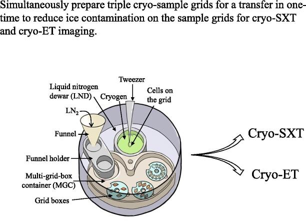
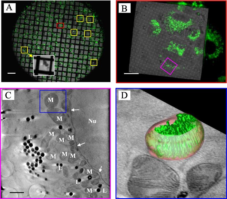
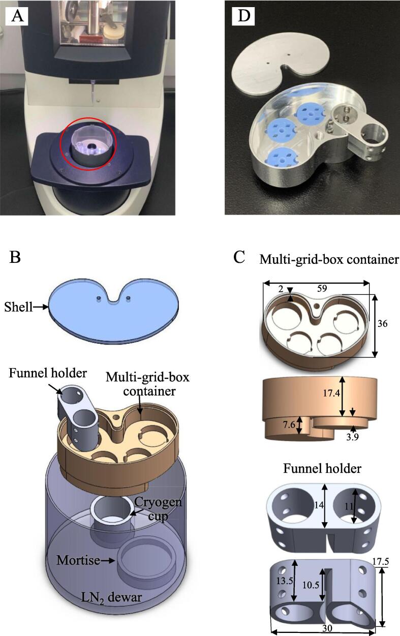
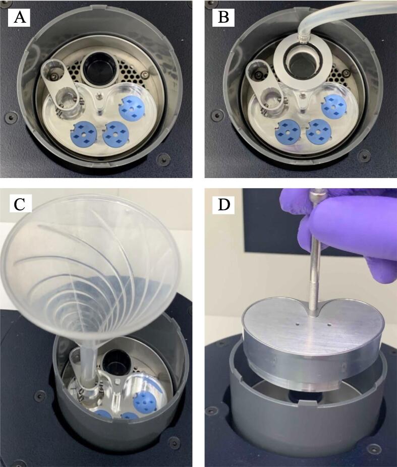
Cryo-soft X-ray tomography (cryo-SXT) is a newly developed technique for imaging 3D whole cells in nearly native states. Cryo-SXT users require the preparation of numerous cryo-sample grids to use the allocated beamtime to study cellular phenomena under various conditions. Therefore, it is important to promptly prepare cryo-sample grids as efficiently and carefully as possible to minimize ice contamination on the frozen sample grid. In this study, we designed a cryo-multi-grid-box storage system, which includes a shell, funnel holder, and multi-grid-box container. Our system not only increases the number of cryo-sample grids that can be temporarily stored but also reduces the frequency of cryo grid-box container transfers, thus decreasing the probability of forming ice on the grid. We have also applied this system to A549 cryo cell grid preparation. The correlative images from cryo-light microscopy and cryo-SXT showed that limited ice had formed on the grid when preparation was performed using our system. Additionally, 3D images of mitochondria with the lamellar shape of the cristae could be observed in our cryo-SXT results. Our cryo-multi-grid-box storage system can be used for cryo-SXT and cryo-electron tomography (cryo-ET) applications.

摘要

低温软X射线断层扫描(cryo-SXT)是一种新开发的用于对近乎天然状态的三维全细胞进行成像的技术。cryo-SXT的用户需要制备大量的低温样品网格,以便利用分配的束流时间来研究各种条件下的细胞现象。因此,尽可能高效且仔细地迅速制备低温样品网格,以尽量减少冷冻样品网格上的冰污染,这一点很重要。在本研究中,我们设计了一种低温多网格盒存储系统,它包括一个外壳、漏斗支架和多网格盒容器。我们的系统不仅增加了可临时存储的低温样品网格数量,还减少了低温网格盒容器的转移频率,从而降低了网格上结冰的概率。我们还将该系统应用于A549细胞低温网格制备。低温光学显微镜和cryo-SXT的相关图像显示,使用我们的系统进行制备时,网格上形成的冰有限。此外,在我们的cryo-SXT结果中可以观察到具有板层状嵴的线粒体的三维图像。我们的低温多网格盒存储系统可用于cryo-SXT和低温电子断层扫描(cryo-ET)应用。

https://cdn.ncbi.nlm.nih.gov/pmc/blobs/d23a/11532778/0e55c6b65f8e/gr3.jpg
https://cdn.ncbi.nlm.nih.gov/pmc/blobs/d23a/11532778/32f88d426f85/ga1.jpg
https://cdn.ncbi.nlm.nih.gov/pmc/blobs/d23a/11532778/49868e8c1e26/gr1.jpg
https://cdn.ncbi.nlm.nih.gov/pmc/blobs/d23a/11532778/5a5faede1147/gr2.jpg
https://cdn.ncbi.nlm.nih.gov/pmc/blobs/d23a/11532778/0e55c6b65f8e/gr3.jpg
https://cdn.ncbi.nlm.nih.gov/pmc/blobs/d23a/11532778/32f88d426f85/ga1.jpg
https://cdn.ncbi.nlm.nih.gov/pmc/blobs/d23a/11532778/49868e8c1e26/gr1.jpg
https://cdn.ncbi.nlm.nih.gov/pmc/blobs/d23a/11532778/5a5faede1147/gr2.jpg
https://cdn.ncbi.nlm.nih.gov/pmc/blobs/d23a/11532778/0e55c6b65f8e/gr3.jpg

相似文献

1
Minimizing ice contamination during specimen preparation for cryo-soft X-ray tomography and cryo-electron tomography.在用于低温软X射线断层扫描和低温电子断层扫描的样本制备过程中尽量减少冰污染。
J Struct Biol X. 2024 Oct 18;10:100113. doi: 10.1016/j.yjsbx.2024.100113. eCollection 2024 Dec.
2
Laboratory based correlative cryo-soft X-ray tomography and cryo-fluorescence microscopy.基于实验室的相关低温冷冻软 X 射线断层扫描和低温荧光显微镜。
Methods Cell Biol. 2024;187:293-320. doi: 10.1016/bs.mcb.2024.02.033. Epub 2024 Mar 22.
3
Correlative cryo-fluorescence and cryo-soft X-ray tomography of adherent cells at European synchrotrons.欧洲同步加速器上贴壁细胞的相关低温荧光和低温软X射线断层扫描
Methods Cell Biol. 2014;124:151-78. doi: 10.1016/B978-0-12-801075-4.00008-2.
4
Cryo-soft X-ray tomography as a quantitative three-dimensional tool to model nanoparticle:cell interaction.低温软X射线断层扫描术作为一种用于模拟纳米颗粒与细胞相互作用的定量三维工具。
J Nanobiotechnology. 2016 Mar 3;14:15. doi: 10.1186/s12951-016-0170-4.
5
3D printed cell culture grid holders for improved cellular specimen preparation in cryo-electron microscopy.3D 打印细胞培养网格支架可改善用于低温电子显微镜的细胞标本制备。
J Struct Biol. 2020 Dec 1;212(3):107633. doi: 10.1016/j.jsb.2020.107633. Epub 2020 Sep 25.
6
Cryo-Electron Microscopy Screening Automation across Multiple Grids using Smart Leginon.使用 Smart Leginon 对多个网格进行冷冻电子显微镜筛选自动化
J Vis Exp. 2023 Dec 1(202). doi: 10.3791/66007.
7
Imaging endosomes and autophagosomes in whole mammalian cells using correlative cryo-fluorescence and cryo-soft X-ray microscopy (cryo-CLXM).使用相关低温荧光和低温软X射线显微镜技术(cryo-CLXM)对完整哺乳动物细胞中的内体和自噬体进行成像。
Ultramicroscopy. 2014 Aug;143(100):77-87. doi: 10.1016/j.ultramic.2013.10.006. Epub 2013 Oct 21.
8
Advanced cryo-tomography workflow developments - correlative microscopy, milling automation and cryo-lift-out.先进的冷冻电子断层扫描工作流程发展——相关显微镜技术、铣削自动化和冷冻提取技术。
J Microsc. 2021 Feb;281(2):112-124. doi: 10.1111/jmi.12939. Epub 2020 Jul 2.
9
Light 'Em up: Efficient Screening of Gold Foil Grids in Cryo-EM.点亮它们:冷冻电镜中金箔网格的高效筛选
Front Mol Biosci. 2022 May 27;9:912363. doi: 10.3389/fmolb.2022.912363. eCollection 2022.
10
Tidy up cryo-EM sample grids with 3D printed tools.使用 3D 打印工具整理冷冻电镜样品网格。
J Struct Biol. 2020 Jan 1;209(1):107414. doi: 10.1016/j.jsb.2019.107414. Epub 2019 Nov 4.

本文引用的文献

1
A 3D Cartographic Description of the Cell by Cryo Soft X-ray Tomography.利用冷冻软 X 射线断层摄影术对细胞进行三维绘图描述。
J Vis Exp. 2021 Mar 15(169). doi: 10.3791/62190.
2
Development of Correlative Cryo-soft X-ray Tomography and Stochastic Reconstruction Microscopy. A Study of Cholesterol Crystal Early Formation in Cells.发展相关的冷冻软 X 射线断层扫描和随机重建显微镜。胆固醇晶体在细胞内早期形成的研究。
J Am Chem Soc. 2016 Nov 16;138(45):14931-14940. doi: 10.1021/jacs.6b07584. Epub 2016 Nov 8.
3
Nanoimaging granule dynamics and subcellular structures in activated mast cells using soft X-ray tomography.
使用软 X 射线断层扫描技术对激活的肥大细胞中的颗粒动态和亚细胞结构进行纳米成像。
Sci Rep. 2016 Oct 17;6:34879. doi: 10.1038/srep34879.
4
Intracellular nanoparticles mass quantification by near-edge absorption soft X-ray nanotomography.通过近边吸收软X射线纳米断层扫描对细胞内纳米颗粒进行质量定量分析。
Sci Rep. 2016 Mar 10;6:22354. doi: 10.1038/srep22354.
5
Imaging endosomes and autophagosomes in whole mammalian cells using correlative cryo-fluorescence and cryo-soft X-ray microscopy (cryo-CLXM).使用相关低温荧光和低温软X射线显微镜技术(cryo-CLXM)对完整哺乳动物细胞中的内体和自噬体进行成像。
Ultramicroscopy. 2014 Aug;143(100):77-87. doi: 10.1016/j.ultramic.2013.10.006. Epub 2013 Oct 21.
6
Cryo X-ray microscope with flat sample geometry for correlative fluorescence and nanoscale tomographic imaging.具有平面样品几何形状的 cryo X 射线显微镜,用于相关荧光和纳米尺度断层成像。
J Struct Biol. 2012 Feb;177(2):212-23. doi: 10.1016/j.jsb.2011.12.023. Epub 2012 Jan 18.
7
Correlative VIS-fluorescence and soft X-ray cryo-microscopy/tomography of adherent cells.贴壁细胞的相关 VIS-荧光和软 X 射线冷冻显微断层扫描。
J Struct Biol. 2012 Feb;177(2):193-201. doi: 10.1016/j.jsb.2011.12.012. Epub 2011 Dec 24.
8
Imaging cellular architecture with X-rays.用 X 射线成像细胞结构。
Curr Opin Struct Biol. 2010 Oct;20(5):623-31. doi: 10.1016/j.sbi.2010.08.008. Epub 2010 Sep 24.
9
X-ray tomography of whole cells.全细胞的X射线断层扫描。
Curr Opin Struct Biol. 2005 Oct;15(5):593-600. doi: 10.1016/j.sbi.2005.08.008.
10
Soft X-ray microscopes and their biological applications.软X射线显微镜及其生物学应用。
Q Rev Biophys. 1995 Feb;28(1):33-130. doi: 10.1017/s0033583500003139.