• 文献检索
  • 文档翻译
  • 深度研究
  • 学术资讯
  • Suppr Zotero 插件Zotero 插件
  • 邀请有礼
  • 套餐&价格
  • 历史记录
应用&插件
Suppr Zotero 插件Zotero 插件浏览器插件Mac 客户端Windows 客户端微信小程序
定价
高级版会员购买积分包购买API积分包
服务
文献检索文档翻译深度研究API 文档MCP 服务
关于我们
关于 Suppr公司介绍联系我们用户协议隐私条款
关注我们

Suppr 超能文献

核心技术专利:CN118964589B侵权必究
粤ICP备2023148730 号-1Suppr @ 2026

文献检索

告别复杂PubMed语法,用中文像聊天一样搜索,搜遍4000万医学文献。AI智能推荐,让科研检索更轻松。

立即免费搜索

文件翻译

保留排版,准确专业,支持PDF/Word/PPT等文件格式,支持 12+语言互译。

免费翻译文档

深度研究

AI帮你快速写综述,25分钟生成高质量综述,智能提取关键信息,辅助科研写作。

立即免费体验

盆腔侧壁的超声评估:方法学共识意见

Ultrasound assessment of the pelvic sidewall: methodological consensus opinion.

作者信息

Fischerova D, Culcasi C, Gatti E, Ng Z, Burgetova A, Szabó G

机构信息

Gynecologic Oncology Centre, Department of Gynecology, Obstetrics and Neonatology, First Faculty of Medicine, Charles University and General University Hospital in Prague, Prague, Czech Republic.

Dipartimento Scienze della Vita e Sanità Pubblica, Università Cattolica del Sacro Cuore, Rome, Italy.

出版信息

Ultrasound Obstet Gynecol. 2025 Jan;65(1):94-105. doi: 10.1002/uog.29122. Epub 2024 Nov 5.

DOI:10.1002/uog.29122
PMID:39499650
原文链接:https://pmc.ncbi.nlm.nih.gov/articles/PMC11693842/
Abstract

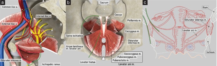
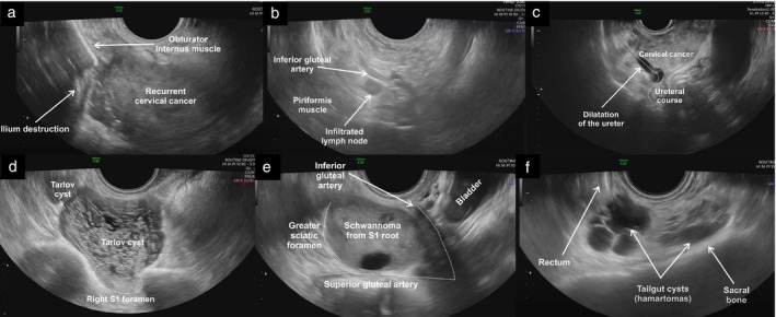
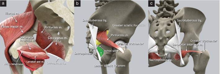
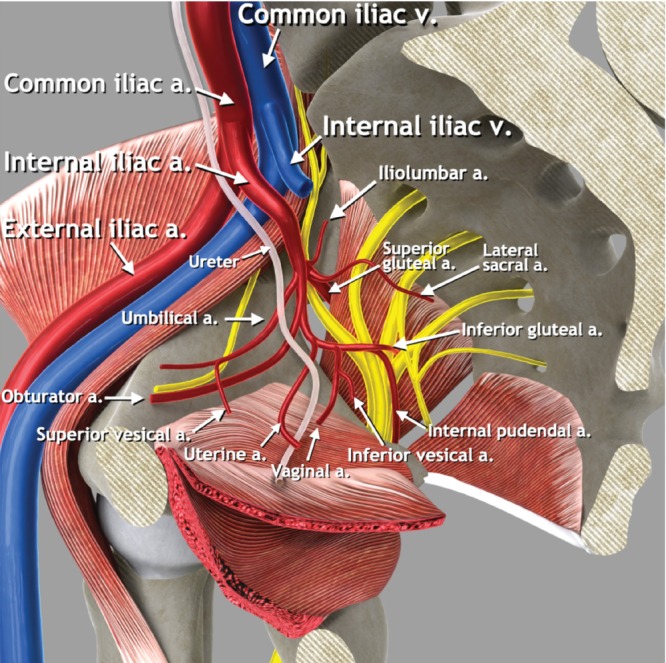
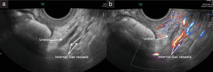
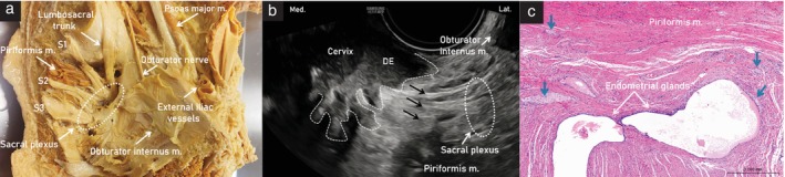
A standardized methodology for the ultrasound evaluation of the pelvic sidewall has not been proposed to date. Herein, a collaborative group of gynecologists and gynecological oncologists with extensive ultrasound experience presents a systematic methodology for the ultrasonographic evaluation of structures within the pelvic sidewall. Five categories of anatomical structures are described (muscles, vessels, lymph nodes, nerves and ureters). A step-by-step transvaginal ultrasound (or, when this is not feasible, transrectal ultrasound) approach is outlined for the evaluation of each anatomical landmark within these categories. Accurate assessment of the pelvic sidewall using a standardized approach improves the detection and diagnosis of non-gynecological pathologies that may mimic gynecological tumors, reducing the risk of unnecessary and even harmful intervention. Furthermore, it plays an important role in completing the staging of malignant gynecological conditions. Transvaginal or transrectal ultrasound therefore represents a viable alternative to magnetic resonance imaging in the preoperative evaluation of lesions affecting the pelvic sidewall, if performed by an expert sonographer. A series of videoclips showing normal and abnormal findings within each respective category illustrates how establishing a universally applicable approach for evaluating this crucial region will be helpful for assessing both benign and malignant conditions affecting the pelvic sidewall. © 2024 The Author(s). Ultrasound in Obstetrics & Gynecology published by John Wiley & Sons Ltd on behalf of International Society of Ultrasound in Obstetrics and Gynecology.

摘要

迄今为止,尚未提出用于盆腔侧壁超声评估的标准化方法。在此,一个由具有丰富超声经验的妇科医生和妇科肿瘤学家组成的协作小组提出了一种用于盆腔侧壁内结构超声评估的系统方法。描述了五类解剖结构(肌肉、血管、淋巴结、神经和输尿管)。概述了一种逐步经阴道超声(或者在不可行时,经直肠超声)方法,用于评估这些类别中的每个解剖标志。使用标准化方法准确评估盆腔侧壁可提高对可能模仿妇科肿瘤的非妇科病变的检测和诊断,降低不必要甚至有害干预的风险。此外,它在完成恶性妇科疾病的分期中起着重要作用。因此,如果由专业超声检查人员进行,经阴道或经直肠超声在影响盆腔侧壁病变的术前评估中是磁共振成像的可行替代方法。一系列显示每个类别中正常和异常发现的视频片段说明了建立一种普遍适用的评估这一关键区域的方法将如何有助于评估影响盆腔侧壁的良性和恶性疾病。© 2024作者。《妇产科超声》由约翰·威利父子有限公司代表国际妇产科超声学会出版。

https://cdn.ncbi.nlm.nih.gov/pmc/blobs/98d5/11693842/2d12fc76dbf7/UOG-65-94-g007.jpg
https://cdn.ncbi.nlm.nih.gov/pmc/blobs/98d5/11693842/07c9d5b0c11e/UOG-65-94-g002.jpg
https://cdn.ncbi.nlm.nih.gov/pmc/blobs/98d5/11693842/0e2e4fd7718f/UOG-65-94-g004.jpg
https://cdn.ncbi.nlm.nih.gov/pmc/blobs/98d5/11693842/4863fc74fe33/UOG-65-94-g005.jpg
https://cdn.ncbi.nlm.nih.gov/pmc/blobs/98d5/11693842/815de73d4a15/UOG-65-94-g010.jpg
https://cdn.ncbi.nlm.nih.gov/pmc/blobs/98d5/11693842/0201e22a0d95/UOG-65-94-g006.jpg
https://cdn.ncbi.nlm.nih.gov/pmc/blobs/98d5/11693842/c8be94b908ba/UOG-65-94-g009.jpg
https://cdn.ncbi.nlm.nih.gov/pmc/blobs/98d5/11693842/99ec05255672/UOG-65-94-g008.jpg
https://cdn.ncbi.nlm.nih.gov/pmc/blobs/98d5/11693842/99e773cb0784/UOG-65-94-g001.jpg
https://cdn.ncbi.nlm.nih.gov/pmc/blobs/98d5/11693842/5f8748042352/UOG-65-94-g003.jpg
https://cdn.ncbi.nlm.nih.gov/pmc/blobs/98d5/11693842/8c32cfabccf7/UOG-65-94-g012.jpg
https://cdn.ncbi.nlm.nih.gov/pmc/blobs/98d5/11693842/7e61834dcedf/UOG-65-94-g011.jpg
https://cdn.ncbi.nlm.nih.gov/pmc/blobs/98d5/11693842/2d12fc76dbf7/UOG-65-94-g007.jpg

相似文献

1
Ultrasound assessment of the pelvic sidewall: methodological consensus opinion.盆腔侧壁的超声评估:方法学共识意见
Ultrasound Obstet Gynecol. 2025 Jan;65(1):94-105. doi: 10.1002/uog.29122. Epub 2024 Nov 5.
2
Ultrasound assessment of lymph nodes for staging of gynecological cancer: consensus opinion on terminology and examination technique.超声评估妇科癌症分期的淋巴结:术语和检查技术的共识意见
Ultrasound Obstet Gynecol. 2025 Feb;65(2):206-225. doi: 10.1002/uog.29127. Epub 2024 Nov 8.
3
Ultrasound examiners' ability to describe ovarian cancer spread using preacquired ultrasound videoclips from a selected patient sample with high prevalence of cancer spread.超声检查人员利用预先获取的、来自癌症扩散患病率高的特定患者样本的超声视频片段来描述卵巢癌扩散情况的能力。
Ultrasound Obstet Gynecol. 2025 May;65(5):641-652. doi: 10.1002/uog.29208. Epub 2025 Apr 18.
4
Learning curve for detection of pelvic parts of ureters by transvaginal sonography: feasibility study.经阴道超声探测输尿管盆段的学习曲线:可行性研究。
Ultrasound Obstet Gynecol. 2020 Feb;55(2):264-268. doi: 10.1002/uog.20379.
5
Magnetic resonance imaging and ultrasound examination in preoperative pelvic staging of early-stage cervical cancer: post-hoc analysis of SENTIX study.磁共振成像和超声检查在早期宫颈癌术前盆腔分期中的应用:SENTIX研究的事后分析
Ultrasound Obstet Gynecol. 2025 Apr;65(4):495-502. doi: 10.1002/uog.29205. Epub 2025 Mar 25.
6
Role of ultrasound in detection of lymph-node metastasis in gynecological cancer: systematic review and meta-analysis.超声在妇科恶性肿瘤淋巴结转移检测中的作用:系统评价和荟萃分析。
Ultrasound Obstet Gynecol. 2024 Aug;64(2):155-163. doi: 10.1002/uog.27633. Epub 2024 Jun 22.
7
Sonographic Evaluation for Endometriosis in Routine Pelvic Ultrasound.经阴道超声常规检查中的子宫内膜异位症的超声评估。
J Minim Invasive Gynecol. 2020 Feb;27(2):265-266. doi: 10.1016/j.jmig.2019.08.027. Epub 2019 Sep 4.
8
Imaging in gynecological disease (26): clinical and ultrasound characteristics of benign retroperitoneal pelvic peripheral-nerve-sheath tumors.妇科疾病影像学(二十六):良性腹膜后盆腔外周神经鞘瘤的临床及超声特征。
Ultrasound Obstet Gynecol. 2023 Nov;62(5):727-738. doi: 10.1002/uog.26223.
9
Vesicoureteral Reflux膀胱输尿管反流
10
Clinical utility of ultrasonography in pediatric and adolescent gynecology: retrospective review of 1313 ultrasound examinations.超声检查在儿科及青少年妇科中的临床应用:1313例超声检查的回顾性分析
Ultrasound Obstet Gynecol. 2025 Feb;65(2):226-234. doi: 10.1002/uog.29155.

引用本文的文献

1
Ultrasound assessment of lymph nodes for staging of gynecological cancer: consensus opinion on terminology and examination technique.超声评估妇科癌症分期的淋巴结:术语和检查技术的共识意见
Ultrasound Obstet Gynecol. 2025 Feb;65(2):206-225. doi: 10.1002/uog.29127. Epub 2024 Nov 8.

本文引用的文献

1
Clinical and ultrasound characteristics of deep endometriosis affecting sacral plexus.影响骶丛的深部子宫内膜异位症的临床和超声特征。
Ultrasound Obstet Gynecol. 2024 Jul;64(1):104-111. doi: 10.1002/uog.27602. Epub 2024 Jun 15.
2
Addendum to consensus opinion from International Deep Endometriosis Analysis (IDEA) group: sonographic evaluation of the parametrium.国际深部子宫内膜异位症分析(IDEA)组共识意见的增编:子宫旁的超声评估。
Ultrasound Obstet Gynecol. 2024 Aug;64(2):275-280. doi: 10.1002/uog.27558. Epub 2024 Jun 30.
3
Dynamic transvaginal ultrasound examination for assessing anatomy of parametrium.
动态经阴道超声检查用于评估子宫旁组织的解剖结构。
Ultrasound Obstet Gynecol. 2023 Dec;62(6):904-906. doi: 10.1002/uog.26313.
4
Consensus on the Gemelli terminology of surgical anatomy for radical hysterectomy.根治性子宫切除术的 Gemelli 外科解剖学术语共识。
Int J Gynecol Cancer. 2023 Jun 5;33(6):876-881. doi: 10.1136/ijgc-2023-004415.
5
Imaging in gynecological disease (26): clinical and ultrasound characteristics of benign retroperitoneal pelvic peripheral-nerve-sheath tumors.妇科疾病影像学(二十六):良性腹膜后盆腔外周神经鞘瘤的临床及超声特征。
Ultrasound Obstet Gynecol. 2023 Nov;62(5):727-738. doi: 10.1002/uog.26223.
6
Visualization of sacral nerve roots and sacral plexus on gynecological transvaginal ultrasound: feasibility study.经阴道妇科超声显示骶神经根和骶丛的可行性研究。
Ultrasound Obstet Gynecol. 2023 Aug;62(2):290-299. doi: 10.1002/uog.26204.
7
Patterns of Recurrent Disease in Cervical Cancer.宫颈癌复发疾病模式
J Pers Med. 2022 May 6;12(5):755. doi: 10.3390/jpm12050755.
8
Cancer of the cervix uteri: 2021 update.子宫颈癌:2021 年更新。
Int J Gynaecol Obstet. 2021 Oct;155 Suppl 1(Suppl 1):28-44. doi: 10.1002/ijgo.13865.
9
Terms, definitions and measurements to describe sonographic features of lymph nodes: consensus opinion from the Vulvar International Tumor Analysis (VITA) group.描述淋巴结超声特征的术语、定义和测量:外阴国际肿瘤分析(VITA)小组的共识意见。
Ultrasound Obstet Gynecol. 2021 Jun;57(6):861-879. doi: 10.1002/uog.23617.
10
Gross and Histologic Anatomy of the Pelvic Ureter: Clinical Applications to Pelvic Surgery.骨盆输尿管的大体解剖和组织解剖:骨盆手术的临床应用。
Obstet Gynecol. 2019 May;133(5):896-904. doi: 10.1097/AOG.0000000000003221.