Department for Companion Animals and Horses, Clinical Unit of Small Animals Internal Medicine, Dermatology, University of Veterinary Medicine Vienna, Vienna, Austria.
Messerli Research Institute, Department of Interdisciplinary Life Sciences, University of Veterinary Medicine Vienna, Veterinärplatz 1, Vienna, A-1210, Austria.
BMC Vet Res. 2024 Nov 6;20(1):506. doi: 10.1186/s12917-024-04350-y.
Iron-deficiency is associated with increased morbidity and mortality in non-communicable diseases. However, iron parameters are rarely assessed in dogs. Here, we aimed to assess and correlate iron parameters in dogs suffering from Canine Atopic Dermatitis (CAD) compared to non-atopic, healthy dogs.
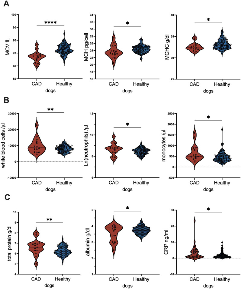
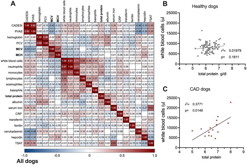
For this retrospective study, blood values and sera of 34 dogs with confirmed CAD were compared with 94 healthy non-atopic dogs. In our cohort, dogs with CAD had significantly lower mean corpuscular volume (MCV, ) mean corpuscular hemoglobin (MCH) but higher white blood cell counts due to increased levels of circulating neutrophils and monocytes. CAD patients also had elevated total protein and c-reactive protein (CRP), but lower albumin levels compared to our healthy control dogs, indicated low-grade inflammation in the CAD cohort. Spearman correlations associated negatively clinical symptom (CADESI-4/PVAS) with MCV; ceruloplasmin and hepcidin, but positively with serum iron. Only in the CAD-cohort, MCV, CRP and albumin-levels negatively affected serum iron-levels and were positively associated with ceruloplasmin. Linear regression analysis revealed that serum iron-levels in CAD subjects, were positively dependent on hematocrit (packed cell volume, PCV) and albumin, and negatively dependent with white blood cells and neutrophils numbers. In contrast, in the healthy cohort, hepcidin was the sole factor associated with serum iron.
A decreased iron status was associated with a higher symptom burden. Iron homeostasis differed markedly in healthy and atopic dermatitis dogs. CAD patients had depleted iron-stores and presented themselves with subclinical inflammation.
铁缺乏与非传染性疾病的发病率和死亡率增加有关。然而,狗的铁参数很少被评估。在这里,我们旨在评估和比较患有犬特应性皮炎(CAD)的狗与非特应性、健康狗的铁参数。
在这项回顾性研究中,我们比较了 34 只确诊为 CAD 的狗的血液值和血清与 94 只健康非特应性狗的血液值和血清。在我们的队列中,患有 CAD 的狗的平均红细胞体积(MCV)和平均红细胞血红蛋白(MCH)明显较低,但由于循环中性粒细胞和单核细胞水平升高,白细胞计数较高。CAD 患者的总蛋白和 C 反应蛋白(CRP)也升高,但与我们的健康对照组相比,白蛋白水平较低,表明 CAD 队列存在低度炎症。Spearman 相关性分析表明,MCV 与临床症状(CADESI-4/PVAS)呈负相关;与血清铁呈负相关,与 ceruloplasmin 和 hepcidin 呈正相关。只有在 CAD 组中,MCV、CRP 和白蛋白水平与血清铁水平呈负相关,与 ceruloplasmin 呈正相关。线性回归分析表明,CAD 患者的血清铁水平与红细胞压积(PCV)和白蛋白呈正相关,与白细胞和中性粒细胞数量呈负相关。相比之下,在健康组中,hepcidin 是唯一与血清铁相关的因素。
铁状态下降与更高的症状负担有关。健康和特应性皮炎犬的铁稳态有明显差异。CAD 患者铁储存减少,并表现出亚临床炎症。