Jayasheelan Shikha, Sreeram Saraswathy, Ns Akash, Mohan Abhay
Department of Pathology, Kasturba Medical College, Mangalore, Manipal Academy of Higher Education, Manipal 576 104, Karnataka, India.
J Surg Case Rep. 2024 Nov 7;2024(11):rjae687. doi: 10.1093/jscr/rjae687. eCollection 2024 Nov.
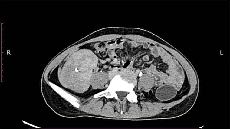
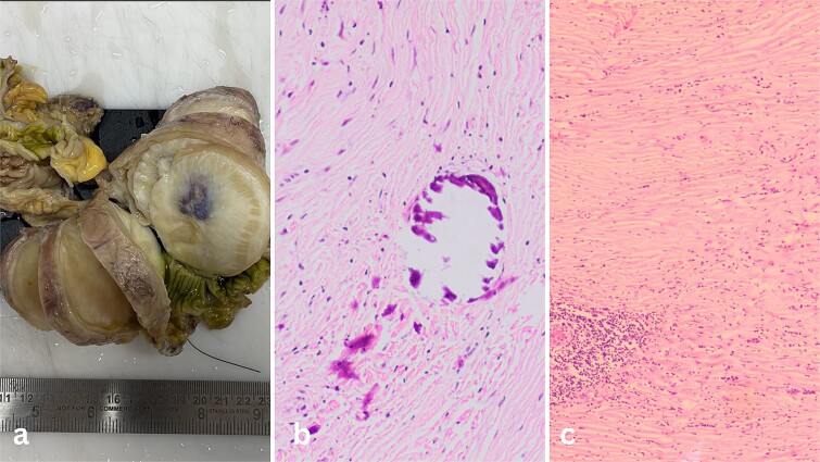
A 19-year-old woman presented with abdominal pain and a palpable mass, initially suspected to be a gastrointestinal stromal tumor (GIST) based on imaging. Surgical excision revealed a sclerotic spindle cell neoplasm with minimal cytological atypia, but immunohistochemistry (IHC) was negative for GIST-specific markers. The pan-negative IHC profile, along with calcification foci and low Ki67 index (<1%), led to a diagnosis of calcifying fibrous tumor (CFT). This case highlights the importance of precise diagnostic evaluation and consideration of rare entities like CFT. Comprehensive histopathological evaluation and IHC are essential diagnostic tools, as they can distinguish between GIST and CFT, leading to accurate treatment and patient management. This case underscores the value of thorough pathological assessment in resolving diagnostic challenges.
一名19岁女性因腹痛和可触及肿块就诊,根据影像学检查最初怀疑为胃肠道间质瘤(GIST)。手术切除显示为硬化性梭形细胞瘤,细胞学异型性极小,但免疫组织化学(IHC)检测GIST特异性标志物呈阴性。免疫组织化学全阴性结果,连同钙化灶和低Ki67指数(<1%),最终诊断为钙化性纤维瘤(CFT)。该病例强调了精确诊断评估以及考虑CFT等罕见实体的重要性。全面的组织病理学评估和免疫组织化学是必不可少的诊断工具,因为它们可以区分GIST和CFT,从而实现准确的治疗和患者管理。该病例强调了全面病理评估在解决诊断难题中的价值。