Department of Radiology, The First Affiliated Hospital, Sun Yat-sen University, Guangzhou, China.
MR Scientific Marketing, Siemens Healthineers Ltd. Guangzhou, China.
Int J Med Sci. 2024 Oct 28;21(14):2799-2806. doi: 10.7150/ijms.104542. eCollection 2024.
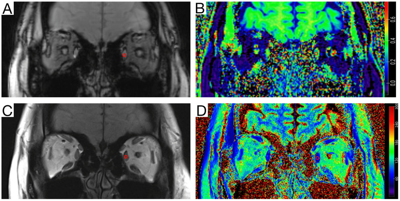
Myasthenia gravis (MG) is an autoimmune neuromuscular disorder that most frequently affects the extraocular muscles (EOMs), which causes symptoms such as ptosis and restricted eye movement. The EOMs in MG patients are representative of autoimmune inflammatory changes in muscle tissue. Currently, there is no reliable, and sensitive imaging technique for monitoring EOM changes to assist in the evaluation of underlying pathological changes. This study included MG patients treated between March and November 2022 at the First Affiliated Hospital of Sun Yat-sen University. Healthy controls (matched by age and sex) were included. Participants underwent 3.0 T MRI with magnetization transfer imaging (MTI) and T2-mapping to measure the magnetization transfer ratio (MTR) and T2-mapping values in the superior, inferior, medial, and lateral rectus muscles. Comparisons were made between MG patients and healthy controls, and between MG subgroups with and without ophthalmoparesis. The MTR and T2-mapping values successfully reflected EOM fibrosis and inflammatory edema in MG patients. MG patients showed significantly higher MTR and T2-mapping values in the EOMs compared with healthy controls. MG patients with ophthalmoparesis exhibited a lower MTR but higher T2-mapping value compared with those without ophthalmoparesis. Combined MTR and T2-mapping values effectively distinguished between MG patients and healthy controls, and between different severities of EOM involvement, with a superior diagnostic accuracy compared with each parameter alone. The combination of MTI and T2-mapping MRI techniques can provide key insight into the pathological changes in EOMs in MG patients. This approach enhances early diagnosis and treatment planning, and therefore may improve clinical outcomes.
重症肌无力(MG)是一种常见的自身免疫性神经肌肉疾病,主要影响眼外肌(EOMs),导致上睑下垂和眼球运动受限等症状。MG 患者的 EOM 表现为肌肉组织的自身免疫性炎症改变。目前,尚无可靠且敏感的成像技术来监测 EOM 变化,以辅助评估潜在的病理变化。
本研究纳入了 2022 年 3 月至 11 月在中山大学第一附属医院接受治疗的 MG 患者。同时纳入了健康对照者(按年龄和性别匹配)。所有参与者均接受了 3.0T MRI 检查,包括磁化传递成像(MTI)和 T2 映射,以测量上直肌、下直肌、内直肌和外直肌的磁化传递率(MTR)和 T2 映射值。比较了 MG 患者与健康对照组、有无眼肌麻痹的 MG 亚组之间的差异。
MTR 和 T2 映射值成功反映了 MG 患者 EOM 的纤维化和炎症性水肿。与健康对照组相比,MG 患者的 EOM 中 MTR 和 T2 映射值显著升高。伴有眼肌麻痹的 MG 患者的 MTR 较低,但 T2 映射值较高。与单独使用每个参数相比,MTR 和 T2 映射值的组合能更有效地区分 MG 患者和健康对照组,以及 EOM 受累的不同严重程度,具有更高的诊断准确性。
MTI 和 T2 映射 MRI 技术的结合可以深入了解 MG 患者 EOM 的病理变化。这种方法可以增强早期诊断和治疗计划,从而改善临床预后。