• 文献检索
  • 文档翻译
  • 深度研究
  • 学术资讯
  • Suppr Zotero 插件Zotero 插件
  • 邀请有礼
  • 套餐&价格
  • 历史记录
应用&插件
Suppr Zotero 插件Zotero 插件浏览器插件Mac 客户端Windows 客户端微信小程序
定价
高级版会员购买积分包购买API积分包
服务
文献检索文档翻译深度研究API 文档MCP 服务
关于我们
关于 Suppr公司介绍联系我们用户协议隐私条款
关注我们

Suppr 超能文献

核心技术专利:CN118964589B侵权必究
粤ICP备2023148730 号-1Suppr @ 2026

文献检索

告别复杂PubMed语法,用中文像聊天一样搜索,搜遍4000万医学文献。AI智能推荐,让科研检索更轻松。

立即免费搜索

文件翻译

保留排版,准确专业,支持PDF/Word/PPT等文件格式,支持 12+语言互译。

免费翻译文档

深度研究

AI帮你快速写综述,25分钟生成高质量综述,智能提取关键信息,辅助科研写作。

立即免费体验

脑胶质母细胞瘤大小和形状的预后意义。

The prognostic importance of glioblastoma size and shape.

机构信息

Department of Neuromedicine and Movement Science, Faculty of Medicine and Health Sciences, Norwegian University of Science and Technology, Trondheim, Norway.

Department of Neuromedicine and Movement Science, Norwegian University of Science and Technology, Trondheim, Norway.

出版信息

Acta Neurochir (Wien). 2024 Nov 12;166(1):450. doi: 10.1007/s00701-024-06351-0.

DOI:10.1007/s00701-024-06351-0
PMID:39531176
原文链接:https://pmc.ncbi.nlm.nih.gov/articles/PMC11557614/
Abstract

PURPOSE

Extent of resection, MGMT promoter methylation status, age, functional level, and residual tumor volume are established prognostic factors for overall survival in glioblastoma patients. Preoperative tumor volume has also been investigated, but the results have been inconclusive. We hypothesized that the surface area and the shape were more representative of the tumor's infiltrative capacities, and thus, the purpose of this study was to assess the prognostic value of tumor size and shape in patients with glioblastoma.

METHODS

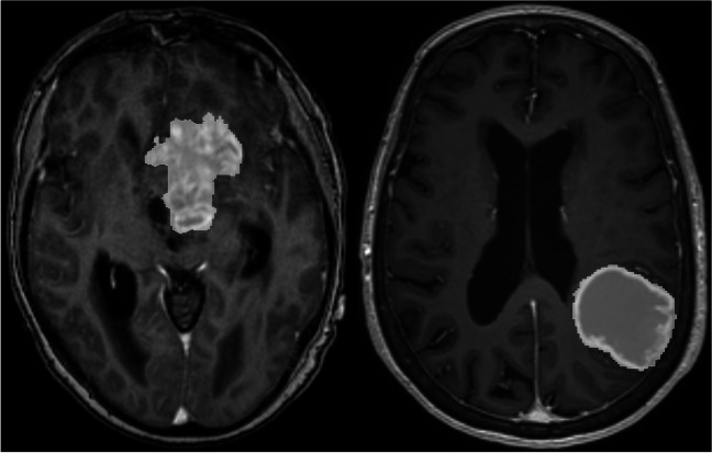
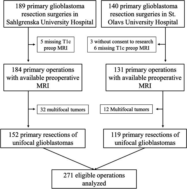
In total, 271 patients with primary, unifocal glioblastoma were included from two centers in Norway and Sweden, respectively. All tumors were automatically segmented on preoperative MRI scans and manually validated. Tumor volume was used as a measurement of size, whereas sphericity index and area-to-volume ratio defined the shape complexity of the tumor. Contact surface area of the tumor was considered a measurement of both size and shape. Multivariable Cox proportional hazards models were used to assess the prognostic value of the respective tumor measurements, with previously established prognostic factors as covariates.

RESULTS

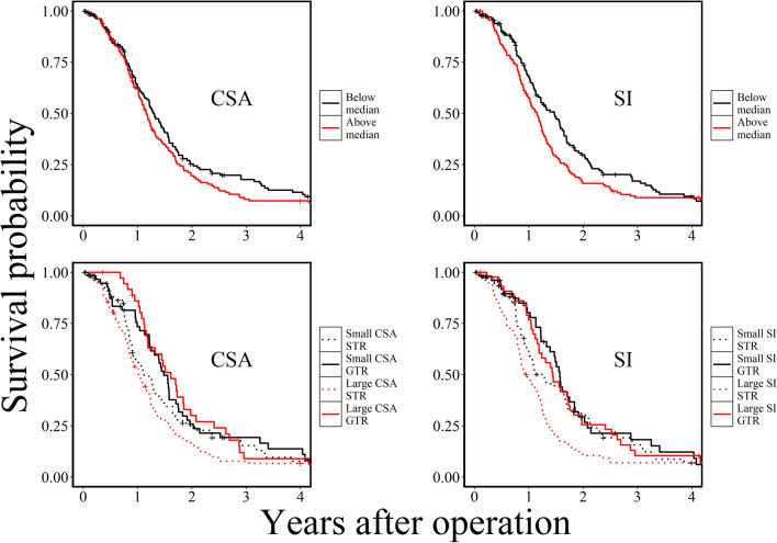
There were no associations between preoperative tumor volume and overall survival. Contact surface area (HR = 1.013, p = 0.002) and sphericity index (HR = 2.223, p = 0.001) were both significant independent prognostic factors for survival in the multivariable Cox models. Contact surface area was also associated with MGMT promoter methylation (p = 0.039) and extent of resection (p = 0.017).

CONCLUSION

Tumor shape complexity appears to be an independent prognostic factor in glioblastoma patients and may also be associated with MGMT promoter methylation status and extent of surgical resection.

摘要

目的

在胶质母细胞瘤患者中,切除范围、MGMT 启动子甲基化状态、年龄、功能状态和残余肿瘤体积是总体生存的既定预后因素。术前肿瘤体积也进行了研究,但结果尚无定论。我们假设肿瘤的表面积和形状更能代表肿瘤的浸润能力,因此,本研究旨在评估胶质母细胞瘤患者肿瘤大小和形状的预后价值。

方法

本研究共纳入来自挪威和瑞典的两个中心的 271 例原发性、单发胶质母细胞瘤患者。所有肿瘤均在术前 MRI 扫描上自动分割,并进行手动验证。肿瘤体积用于测量大小,而球形指数和面积-体积比定义了肿瘤形状的复杂性。肿瘤的接触表面积被认为是大小和形状的测量指标。多变量 Cox 比例风险模型用于评估各自肿瘤测量的预后价值,同时将先前确定的预后因素作为协变量。

结果

术前肿瘤体积与总生存期之间无相关性。接触表面积(HR=1.013,p=0.002)和球形指数(HR=2.223,p=0.001)均为多变量 Cox 模型中生存的独立显著预后因素。接触表面积也与 MGMT 启动子甲基化(p=0.039)和切除范围(p=0.017)相关。

结论

肿瘤形状的复杂性似乎是胶质母细胞瘤患者的独立预后因素,并且可能与 MGMT 启动子甲基化状态和手术切除范围相关。

https://cdn.ncbi.nlm.nih.gov/pmc/blobs/f8f2/11557614/a0d91dbcbba0/701_2024_6351_Fig3_HTML.jpg
https://cdn.ncbi.nlm.nih.gov/pmc/blobs/f8f2/11557614/ae64caf2be50/701_2024_6351_Fig1_HTML.jpg
https://cdn.ncbi.nlm.nih.gov/pmc/blobs/f8f2/11557614/bc2be0298958/701_2024_6351_Fig2_HTML.jpg
https://cdn.ncbi.nlm.nih.gov/pmc/blobs/f8f2/11557614/a0d91dbcbba0/701_2024_6351_Fig3_HTML.jpg
https://cdn.ncbi.nlm.nih.gov/pmc/blobs/f8f2/11557614/ae64caf2be50/701_2024_6351_Fig1_HTML.jpg
https://cdn.ncbi.nlm.nih.gov/pmc/blobs/f8f2/11557614/bc2be0298958/701_2024_6351_Fig2_HTML.jpg
https://cdn.ncbi.nlm.nih.gov/pmc/blobs/f8f2/11557614/a0d91dbcbba0/701_2024_6351_Fig3_HTML.jpg

相似文献

1
The prognostic importance of glioblastoma size and shape.脑胶质母细胞瘤大小和形状的预后意义。
Acta Neurochir (Wien). 2024 Nov 12;166(1):450. doi: 10.1007/s00701-024-06351-0.
2
Systemic treatments for metastatic cutaneous melanoma.转移性皮肤黑色素瘤的全身治疗
Cochrane Database Syst Rev. 2018 Feb 6;2(2):CD011123. doi: 10.1002/14651858.CD011123.pub2.
3
Cost-effectiveness of using prognostic information to select women with breast cancer for adjuvant systemic therapy.利用预后信息为乳腺癌患者选择辅助性全身治疗的成本效益
Health Technol Assess. 2006 Sep;10(34):iii-iv, ix-xi, 1-204. doi: 10.3310/hta10340.
4
Impact of residual disease as a prognostic factor for survival in women with advanced epithelial ovarian cancer after primary surgery.原发性手术后晚期上皮性卵巢癌患者残留病灶对生存预后的影响。
Cochrane Database Syst Rev. 2022 Sep 26;9(9):CD015048. doi: 10.1002/14651858.CD015048.pub2.
5
Treatment options for progression or recurrence of glioblastoma: a network meta-analysis.治疗胶质母细胞瘤进展或复发的选择:网络荟萃分析。
Cochrane Database Syst Rev. 2021 May 4;5(1):CD013579. doi: 10.1002/14651858.CD013579.pub2.
6
Molecular Biomarkers in Glioblastoma: A Systematic Review and Meta-Analysis.胶质母细胞瘤中的分子生物标志物:系统评价和荟萃分析。
Int J Mol Sci. 2022 Aug 9;23(16):8835. doi: 10.3390/ijms23168835.
7
Intraoperative imaging technology to maximise extent of resection for glioma.术中成像技术以最大化胶质瘤的切除范围。
Cochrane Database Syst Rev. 2018 Jan 22;1(1):CD012788. doi: 10.1002/14651858.CD012788.pub2.
8
MGMT methylation and its prognostic significance in inoperable IDH-wildtype glioblastoma: the MGMT-GBM study.MGMT 甲基化及其在不可切除 IDH 野生型胶质母细胞瘤中的预后意义:MGMT-GBM 研究。
Acta Neurochir (Wien). 2024 Oct 5;166(1):394. doi: 10.1007/s00701-024-06300-x.
9
Magnetic resonance perfusion for differentiating low-grade from high-grade gliomas at first presentation.首次就诊时磁共振灌注成像用于鉴别低级别与高级别胶质瘤
Cochrane Database Syst Rev. 2018 Jan 22;1(1):CD011551. doi: 10.1002/14651858.CD011551.pub2.
10
Home treatment for mental health problems: a systematic review.心理健康问题的居家治疗:一项系统综述
Health Technol Assess. 2001;5(15):1-139. doi: 10.3310/hta5150.

引用本文的文献

1
Survival prediction with radiomics for patients with IDH mutated lower-grade glioma.基于影像组学的异柠檬酸脱氢酶(IDH)突变型低级别胶质瘤患者生存预测
J Neurooncol. 2025 Mar 18. doi: 10.1007/s11060-025-05006-z.

本文引用的文献

1
Segmentation of glioblastomas in early post-operative multi-modal MRI with deep neural networks.基于深度神经网络的多模态 MRI 术后早期胶质母细胞瘤分割。
Sci Rep. 2023 Nov 2;13(1):18897. doi: 10.1038/s41598-023-45456-x.
2
Beyond Imaging and Genetic Signature in Glioblastoma: Radiogenomic Holistic Approach in Neuro-Oncology.胶质母细胞瘤中超越影像学和基因特征:神经肿瘤学中的放射基因组整体方法
Biomedicines. 2022 Dec 9;10(12):3205. doi: 10.3390/biomedicines10123205.
3
Preoperative Brain Tumor Imaging: Models and Software for Segmentation and Standardized Reporting.
术前脑肿瘤成像:用于分割和标准化报告的模型与软件
Front Neurol. 2022 Jul 27;13:932219. doi: 10.3389/fneur.2022.932219. eCollection 2022.
4
Prognostic validation of a new classification system for extent of resection in glioblastoma: A report of the RANO resect group.新胶质母细胞瘤切除范围分类系统的预后验证: RANO 切除组报告。
Neuro Oncol. 2023 May 4;25(5):940-954. doi: 10.1093/neuonc/noac193.
5
Epidemiology of Glioblastoma Multiforme-Literature Review.多形性胶质母细胞瘤的流行病学——文献综述
Cancers (Basel). 2022 May 13;14(10):2412. doi: 10.3390/cancers14102412.
6
Volumetric assessment of glioblastoma and its predictive value for survival.脑胶质母细胞瘤的体积评估及其对生存的预测价值。
Acta Neurochir (Wien). 2019 Aug;161(8):1723-1732. doi: 10.1007/s00701-019-03966-6. Epub 2019 Jun 28.
7
Osseous versus Nonosseous Spinal Epidural Arteriovenous Fistulas: Experiences of 13 Patients.骨内型与非骨内型硬脊膜动静脉瘘:13 例患者的经验。
AJNR Am J Neuroradiol. 2019 Jan;40(1):129-134. doi: 10.3174/ajnr.A5904. Epub 2018 Dec 6.
8
An Experimenter's Guide to Glioblastoma Invasion Pathways.胶质母细胞瘤侵袭途径实验者指南。
Trends Mol Med. 2018 Sep;24(9):763-780. doi: 10.1016/j.molmed.2018.07.003. Epub 2018 Jul 30.
9
Clinically Relevant Imaging Features for Promoter Methylation in Multiple Glioblastoma Studies: A Systematic Review and Meta-Analysis.多个胶质母细胞瘤研究中启动子甲基化的临床相关影像学特征:系统评价和荟萃分析。
AJNR Am J Neuroradiol. 2018 Aug;39(8):1439-1445. doi: 10.3174/ajnr.A5711. Epub 2018 Jul 12.
10
Time-varying covariates and coefficients in Cox regression models.Cox回归模型中的时变协变量和系数。
Ann Transl Med. 2018 Apr;6(7):121. doi: 10.21037/atm.2018.02.12.