• 文献检索
  • 文档翻译
  • 深度研究
  • 学术资讯
  • Suppr Zotero 插件Zotero 插件
  • 邀请有礼
  • 套餐&价格
  • 历史记录
应用&插件
Suppr Zotero 插件Zotero 插件浏览器插件Mac 客户端Windows 客户端微信小程序
定价
高级版会员购买积分包购买API积分包
服务
文献检索文档翻译深度研究API 文档MCP 服务
关于我们
关于 Suppr公司介绍联系我们用户协议隐私条款
关注我们

Suppr 超能文献

核心技术专利:CN118964589B侵权必究
粤ICP备2023148730 号-1Suppr @ 2026

文献检索

告别复杂PubMed语法,用中文像聊天一样搜索,搜遍4000万医学文献。AI智能推荐,让科研检索更轻松。

立即免费搜索

文件翻译

保留排版,准确专业,支持PDF/Word/PPT等文件格式,支持 12+语言互译。

免费翻译文档

深度研究

AI帮你快速写综述,25分钟生成高质量综述,智能提取关键信息,辅助科研写作。

立即免费体验

皮肤 T 细胞淋巴瘤图谱揭示了恶性 T2 细胞,这些细胞由富含 B 细胞的肿瘤微环境支持。

Cutaneous T cell lymphoma atlas reveals malignant T2 cells supported by a B cell-rich tumor microenvironment.

机构信息

Wellcome Sanger Institute, Wellcome Genome Campus, Hinxton, UK.

Department of Systems Biology, The University of Texas MD Anderson Cancer Center, Houston, TX, USA.

出版信息

Nat Immunol. 2024 Dec;25(12):2320-2330. doi: 10.1038/s41590-024-02018-1. Epub 2024 Nov 18.

DOI:10.1038/s41590-024-02018-1
PMID:39558094
原文链接:https://pmc.ncbi.nlm.nih.gov/articles/PMC11588665/
Abstract

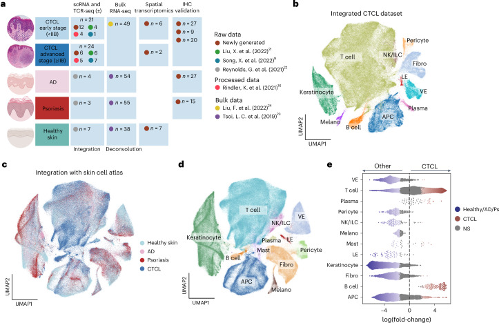
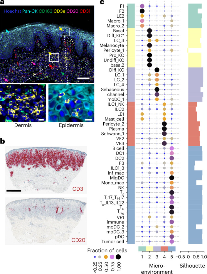
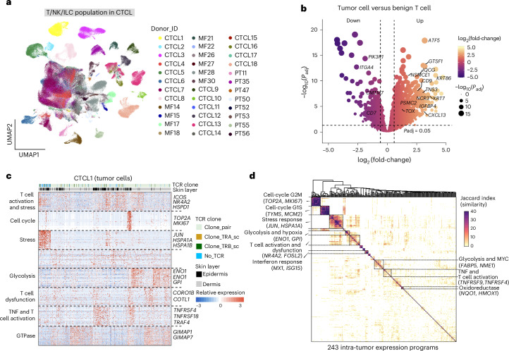
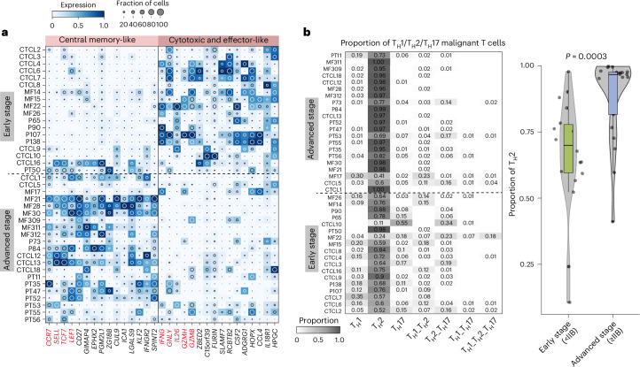
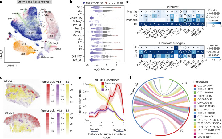
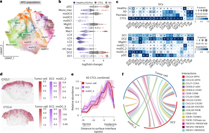
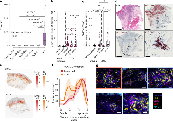
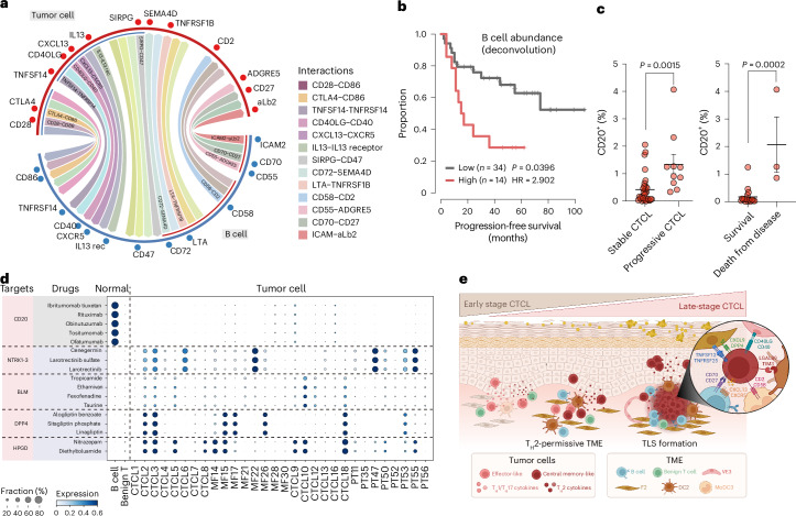
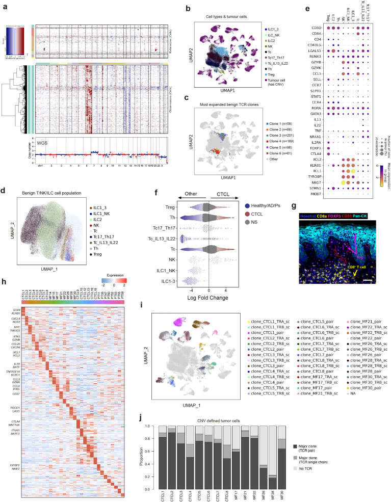
Cutaneous T cell lymphoma (CTCL) is a potentially fatal clonal malignancy of T cells primarily affecting the skin. The most common form of CTCL, mycosis fungoides, can be difficult to diagnose, resulting in treatment delay. We performed single-cell and spatial transcriptomics analysis of skin from patients with mycosis fungoides-type CTCL and an integrated comparative analysis with human skin cell atlas datasets from healthy and inflamed skin. We revealed the co-optation of T helper 2 (T2) cell-immune gene programs by malignant CTCL cells and modeling of the tumor microenvironment to support their survival. We identified MHC-II fibroblasts and dendritic cells that can maintain T2 cell-like tumor cells. CTCL tumor cells are spatially associated with B cells, forming tertiary lymphoid structure-like aggregates. Finally, we validated the enrichment of B cells in CTCL and its association with disease progression across three independent patient cohorts. Our findings provide diagnostic aids, potential biomarkers for disease staging and therapeutic strategies for CTCL.

摘要

皮肤 T 细胞淋巴瘤 (CTCL) 是一种潜在致命的 T 细胞克隆性恶性肿瘤,主要影响皮肤。蕈样真菌病是 CTCL 最常见的形式,其诊断较为困难,导致治疗延误。我们对蕈样真菌病型 CTCL 患者的皮肤进行了单细胞和空间转录组学分析,并与健康和炎症皮肤的人类皮肤细胞图谱数据集进行了综合比较分析。我们揭示了恶性 CTCL 细胞对辅助性 T 细胞 2(T2)细胞免疫基因程序的共调控,以及对肿瘤微环境的建模以支持其存活。我们鉴定了 MHC-II 成纤维细胞和树突状细胞,它们可以维持 T2 样肿瘤细胞。CTCL 肿瘤细胞与 B 细胞在空间上相关,形成类似三级淋巴结构的聚集物。最后,我们在三个独立的患者队列中验证了 CTCL 中 B 细胞的富集及其与疾病进展的相关性。我们的研究结果为 CTCL 提供了诊断辅助工具、疾病分期的潜在生物标志物和治疗策略。

https://cdn.ncbi.nlm.nih.gov/pmc/blobs/7c22/11588665/70cbbeac9a8b/41590_2024_2018_Fig14_ESM.jpg
https://cdn.ncbi.nlm.nih.gov/pmc/blobs/7c22/11588665/107c5dac718a/41590_2024_2018_Fig1_HTML.jpg
https://cdn.ncbi.nlm.nih.gov/pmc/blobs/7c22/11588665/0b669eced4e0/41590_2024_2018_Fig2_HTML.jpg
https://cdn.ncbi.nlm.nih.gov/pmc/blobs/7c22/11588665/7d184894c807/41590_2024_2018_Fig3_HTML.jpg
https://cdn.ncbi.nlm.nih.gov/pmc/blobs/7c22/11588665/7f55d5012f0c/41590_2024_2018_Fig4_HTML.jpg
https://cdn.ncbi.nlm.nih.gov/pmc/blobs/7c22/11588665/2a912944638f/41590_2024_2018_Fig5_HTML.jpg
https://cdn.ncbi.nlm.nih.gov/pmc/blobs/7c22/11588665/5999092180e7/41590_2024_2018_Fig6_HTML.jpg
https://cdn.ncbi.nlm.nih.gov/pmc/blobs/7c22/11588665/14b7f09adfbc/41590_2024_2018_Fig7_HTML.jpg
https://cdn.ncbi.nlm.nih.gov/pmc/blobs/7c22/11588665/360f2cc0373c/41590_2024_2018_Fig8_HTML.jpg
https://cdn.ncbi.nlm.nih.gov/pmc/blobs/7c22/11588665/58d2df60555a/41590_2024_2018_Fig9_ESM.jpg
https://cdn.ncbi.nlm.nih.gov/pmc/blobs/7c22/11588665/fe8f4b5c6bc8/41590_2024_2018_Fig10_ESM.jpg
https://cdn.ncbi.nlm.nih.gov/pmc/blobs/7c22/11588665/33c5bc3d4d10/41590_2024_2018_Fig11_ESM.jpg
https://cdn.ncbi.nlm.nih.gov/pmc/blobs/7c22/11588665/a8d1032cca57/41590_2024_2018_Fig12_ESM.jpg
https://cdn.ncbi.nlm.nih.gov/pmc/blobs/7c22/11588665/036991340355/41590_2024_2018_Fig13_ESM.jpg
https://cdn.ncbi.nlm.nih.gov/pmc/blobs/7c22/11588665/70cbbeac9a8b/41590_2024_2018_Fig14_ESM.jpg

相似文献

1
Cutaneous T cell lymphoma atlas reveals malignant T2 cells supported by a B cell-rich tumor microenvironment.皮肤 T 细胞淋巴瘤图谱揭示了恶性 T2 细胞,这些细胞由富含 B 细胞的肿瘤微环境支持。
Nat Immunol. 2024 Dec;25(12):2320-2330. doi: 10.1038/s41590-024-02018-1. Epub 2024 Nov 18.
2
Cutaneous T cell lymphoma: the helping hand of dendritic cells.皮肤T细胞淋巴瘤:树突状细胞的助力
Ann N Y Acad Sci. 2001 Sep;941:1-11.
3
Single-Cell Lymphocyte Heterogeneity in Advanced Cutaneous T-cell Lymphoma Skin Tumors.晚期皮肤 T 细胞淋巴瘤皮肤肿瘤中单细胞淋巴细胞异质性。
Clin Cancer Res. 2019 Jul 15;25(14):4443-4454. doi: 10.1158/1078-0432.CCR-19-0148. Epub 2019 Apr 22.
4
Normal and cancer fibroblasts differentially regulate TWIST1, TOX and cytokine gene expression in cutaneous T-cell lymphoma.正常成纤维细胞和癌细胞在皮肤 T 细胞淋巴瘤中差异调节 TWIST1、TOX 和细胞因子基因的表达。
BMC Cancer. 2021 May 3;21(1):492. doi: 10.1186/s12885-021-08142-7.
5
Low herpesvirus entry mediator (HVEM) expression on dermal fibroblasts contributes to a Th2-dominant microenvironment in advanced cutaneous T-cell lymphoma.真皮成纤维细胞中低水平的疱疹病毒进入介体 (HVEM) 表达导致晚期皮肤 T 细胞淋巴瘤中 Th2 占优势的微环境。
J Invest Dermatol. 2012 Apr;132(4):1280-9. doi: 10.1038/jid.2011.470. Epub 2012 Feb 2.
6
High numbers of DC-SIGN+ dendritic cells in lesional skin of cutaneous T-cell lymphoma.皮肤 T 细胞淋巴瘤皮损中 DC-SIGN+树突状细胞数量较多。
J Am Acad Dermatol. 2010 Jun;62(6):995-1004. doi: 10.1016/j.jaad.2009.06.082.
7
Primary cutaneous T-cell lymphoma (mycosis fungoides and Sézary syndrome): part I. Diagnosis: clinical and histopathologic features and new molecular and biologic markers.原发性皮肤 T 细胞淋巴瘤(蕈样肉芽肿和赛泽里综合征):第一部分。诊断:临床和组织病理学特征以及新的分子和生物学标志物。
J Am Acad Dermatol. 2014 Feb;70(2):205.e1-16; quiz 221-2. doi: 10.1016/j.jaad.2013.07.049.
8
Single-cell RNA sequencing comparison of CD4+, CD8+ and T-cell receptor γδ+ cutaneous T-cell lymphomas reveals subset-specific molecular phenotypes.CD4 +、CD8 +和T细胞受体γδ +皮肤T细胞淋巴瘤的单细胞RNA测序比较揭示了亚群特异性分子表型。
Br J Dermatol. 2025 Jan 24;192(2):269-282. doi: 10.1093/bjd/ljae313.
9
Analysis of STAT4 expression in cutaneous T-cell lymphoma (CTCL) patients and patient-derived cell lines.皮肤T细胞淋巴瘤(CTCL)患者及患者来源细胞系中STAT4表达的分析。
Cell Cycle. 2014;13(18):2975-82. doi: 10.4161/15384101.2014.947759.
10
TH2 cytokines from malignant cells suppress TH1 responses and enforce a global TH2 bias in leukemic cutaneous T-cell lymphoma.恶性细胞产生的 TH2 细胞因子抑制 TH1 反应,并在白血病皮肤 T 细胞淋巴瘤中强制形成全身性 TH2 偏向。
Clin Cancer Res. 2013 Jul 15;19(14):3755-63. doi: 10.1158/1078-0432.CCR-12-3488. Epub 2013 Jun 19.

引用本文的文献

1
An Update on Single-Cell RNA Sequencing in Illuminating Disease Mechanisms of Cutaneous T-Cell Lymphoma.单细胞RNA测序在揭示皮肤T细胞淋巴瘤疾病机制方面的最新进展
Cancers (Basel). 2025 Sep 5;17(17):2921. doi: 10.3390/cancers17172921.
2
Dermatopathological Challenges in Objectively Characterizing Immunotherapy Response in Mycosis Fungoides.蕈样肉芽肿免疫治疗反应客观特征化中的皮肤病理学挑战
Dermatopathology (Basel). 2025 Jul 29;12(3):22. doi: 10.3390/dermatopathology12030022.
3
Skin-Directed Therapies in Mycosis Fungoides: An Update.

本文引用的文献

1
Integrative mapping of human CD8 T cells in inflammation and cancer.人类CD8 T细胞在炎症和癌症中的综合图谱分析。
Nat Methods. 2025 Feb;22(2):435-445. doi: 10.1038/s41592-024-02530-0. Epub 2024 Nov 29.
2
EvoAI enables extreme compression and reconstruction of the protein sequence space.EvoAI可实现蛋白质序列空间的极致压缩与重建。
Nat Methods. 2025 Jan;22(1):102-112. doi: 10.1038/s41592-024-02504-2. Epub 2024 Nov 11.
3
WebAtlas pipeline for integrated single-cell and spatial transcriptomic data.用于整合单细胞和空间转录组数据的WebAtlas工作流程
蕈样肉芽肿的皮肤定向治疗:最新进展
Dermatol Ther (Heidelb). 2025 Aug 14. doi: 10.1007/s13555-025-01511-1.
4
Sketching T cell atlases in the single-cell era: challenges and recommendations.单细胞时代绘制T细胞图谱:挑战与建议
Immunol Cell Biol. 2025 Aug;103(7):723-737. doi: 10.1111/imcb.70040. Epub 2025 Jun 29.
5
Mycosis Fungoides, Sézary Syndrome, and Cutaneous B-Cell Lymphomas: 2025 Update on Diagnosis, Risk-Stratification, and Management.蕈样肉芽肿、塞扎里综合征和皮肤B细胞淋巴瘤:2025年诊断、风险分层及管理的最新进展
Am J Hematol. 2025 Sep;100(9):1603-1628. doi: 10.1002/ajh.27735. Epub 2025 Jun 10.
6
Cutaneous T Cell Lymphoma Following Dupilumab Therapy in Patients with Atopic Dermatitis: Clinical Review and Recommendations.度普利尤单抗治疗特应性皮炎患者后发生的皮肤T细胞淋巴瘤:临床综述与建议
Am J Clin Dermatol. 2025 May 26. doi: 10.1007/s40257-025-00955-7.
7
Activation-induced cytidine deaminase in tertiary lymphoid structures: dual roles and implications in cancer prognosis.三级淋巴结构中的活化诱导胞苷脱氨酶:双重作用及其对癌症预后的影响
Front Oncol. 2025 Apr 9;15:1555491. doi: 10.3389/fonc.2025.1555491. eCollection 2025.
8
Multiplexed imaging mass cytometry elicits p-S6 as a therapeutic and prognostic factor in diffuse large B-cell lymphoma.多重成像质谱流式细胞术揭示p-S6作为弥漫性大B细胞淋巴瘤的治疗和预后因素。
Blood Adv. 2025 Jul 8;9(13):3249-3262. doi: 10.1182/bloodadvances.2024015469.
9
Single-cell profiling uncovers the intricate pathological niche diversity in brain, lymph node, bone, and adrenal metastases of lung cancer.单细胞分析揭示了肺癌脑、淋巴结、骨和肾上腺转移灶中复杂的病理微环境多样性。
Discov Oncol. 2025 Apr 10;16(1):512. doi: 10.1007/s12672-025-02269-w.
Nat Methods. 2025 Jan;22(1):3-5. doi: 10.1038/s41592-024-02371-x.
4
Automatic cell-type harmonization and integration across Human Cell Atlas datasets.自动细胞类型协调和整合人类细胞图谱数据集。
Cell. 2023 Dec 21;186(26):5876-5891.e20. doi: 10.1016/j.cell.2023.11.026.
5
Unsupervised removal of systematic background noise from droplet-based single-cell experiments using CellBender.基于 CellBender 的无监督去除液滴式单细胞实验系统背景噪声。
Nat Methods. 2023 Sep;20(9):1323-1335. doi: 10.1038/s41592-023-01943-7. Epub 2023 Aug 7.
6
Spatially resolved multiomics of human cardiac niches.人类心脏龛位的空间分辨多组学研究。
Nature. 2023 Jul;619(7971):801-810. doi: 10.1038/s41586-023-06311-1. Epub 2023 Jul 12.
7
Hallmarks of transcriptional intratumour heterogeneity across a thousand tumours.一千个肿瘤中的转录肿瘤内异质性特征。
Nature. 2023 Jun;618(7965):598-606. doi: 10.1038/s41586-023-06130-4. Epub 2023 May 31.
8
Diagnosis of mycosis fungoides or Sézary syndrome after dupilumab use: A systematic review.使用度普利尤单抗后蕈样肉芽肿或塞扎里综合征的诊断:一项系统评价
J Am Acad Dermatol. 2023 May;88(5):1164-1166. doi: 10.1016/j.jaad.2022.12.001. Epub 2022 Dec 5.
9
Single-cell RNA sequencing unveils the communications between malignant T and myeloid cells contributing to tumor growth and immunosuppression in cutaneous T-cell lymphoma.单细胞RNA测序揭示了恶性T细胞与髓样细胞之间的通讯,这种通讯促进了皮肤T细胞淋巴瘤的肿瘤生长和免疫抑制。
Cancer Lett. 2022 Dec 28;551:215972. doi: 10.1016/j.canlet.2022.215972. Epub 2022 Oct 17.
10
Cutaneous T-cell lymphoma following dupilumab use: a systematic review.使用度普利尤单抗后发生的皮肤 T 细胞淋巴瘤:系统评价。
Int J Dermatol. 2023 Jul;62(7):862-876. doi: 10.1111/ijd.16388. Epub 2022 Sep 8.