Tan Josephine L, Kalia Vibhuti, Pautler Stephen E, Bauman Glenn, Gast Lena V, Müller Max, Nagel Armin M, Thiessen Jonathan D, Scholl Timothy J, Akbari Alireza
Medical Biophysics, Western University, London, N6A 3K7, Canada.
Medical Imaging, Western University, London, N6A 3K7, Canada.
Radiol Adv. 2024 Sep 30;1(3):umae023. doi: 10.1093/radadv/umae023. eCollection 2024 Sep.
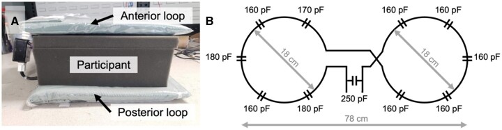
Sodium (Na) MRI of prostate cancer (PCa) is a novel but underdocumented technique conventionally acquired using an endorectal coil. These endorectal coils are associated with challenges (e.g., a nonuniform sensitivity profile, limited prostate coverage, patient discomfort) that could be mitigated with an external Na MRI coil.
To quantify tissue sodium concentration (TSC) differences within the prostate of participants with PCa and healthy volunteers using an external Na MRI radiofrequency coil at 3 T.
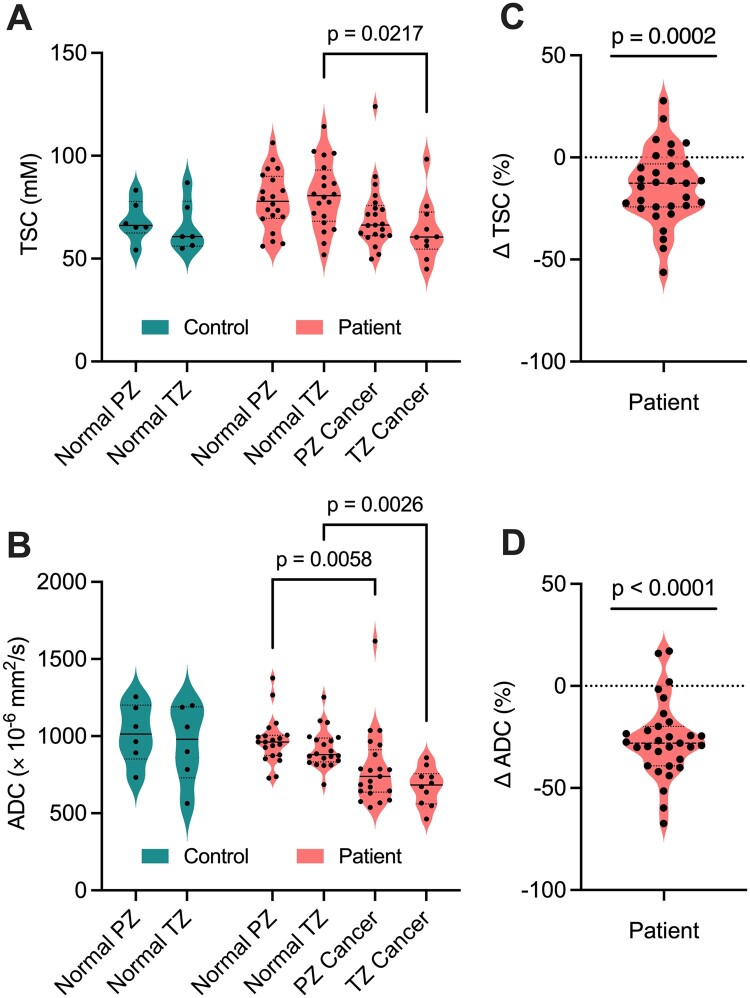
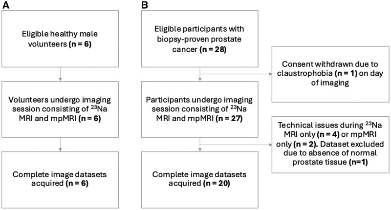
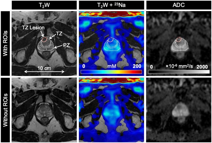
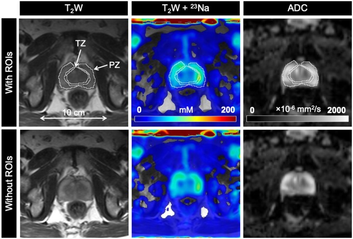
A prospective study was conducted from January 2022 to June 2024 in healthy volunteers and participants with biopsy-proven PCa. Prostate Na MRI was acquired on a 3-T PET/MRI scanner using a custom-built 2-loop (diameter, 18 cm) butterfly surface coil tuned for the Na frequency (32.6 MHz). The percent difference in TSC (ΔTSC) between prostate cancer lesions and surrounding noncancerous prostate tissue of the peripheral zone (PZ) and transition zone (TZ) was evaluated using a 1-sample -test. TSC was compared to apparent diffusion coefficient (ADC) measurements as a clinical reference.
Six healthy volunteers (mean age, 54.5 years ± 12.7) and 20 participants with PCa (mean age, 70.7 years ± 8.3) were evaluated. A total of 31 lesions were detected (21 PZ, 10 TZ) across PCa participants. Compared to noncancerous prostate tissue, prostate cancer lesions had significantly lower TSC (ΔTSC, -14.1% ± 18.2, = .0002) and ADC (ΔADC, -26.6% ± 18.7, < .0001).
We used an external Na MRI coil for whole-gland comparison of TSC in PCa and noncancerous prostate tissue at 3 T. PCa lesions presented with lower TSC compared to surrounding noncancerous PZ and TZ tissue. These findings demonstrate the feasibility of an external Na MRI coil to quantify TSC in the prostate and offer a promising, noninvasive approach to PCa diagnosis and management.
前列腺癌(PCa)的钠(Na)磁共振成像(MRI)是一种新颖但文献记载较少的技术,传统上使用直肠内线圈进行采集。这些直肠内线圈存在一些挑战(例如,灵敏度分布不均匀、前列腺覆盖范围有限、患者不适),而外部Na MRI线圈可以缓解这些问题。
使用3 T的外部Na MRI射频线圈,量化PCa参与者和健康志愿者前列腺内的组织钠浓度(TSC)差异。
2022年1月至2024年6月对健康志愿者和经活检证实患有PCa的参与者进行了一项前瞻性研究。使用定制的2环(直径18 cm)蝶形表面线圈在3 T的PET/MRI扫描仪上采集前列腺Na MRI,该线圈针对Na频率(32.6 MHz)进行了调谐。使用单样本检验评估前列腺癌病变与外周区(PZ)和移行区(TZ)周围非癌性前列腺组织之间TSC的百分比差异(ΔTSC)。将TSC与表观扩散系数(ADC)测量值作为临床参考进行比较。
评估了6名健康志愿者(平均年龄54.5岁±12.7)和20名PCa参与者(平均年龄70.7岁±8.3)。在PCa参与者中总共检测到31个病变(21个在PZ,10个在TZ)。与非癌性前列腺组织相比,前列腺癌病变的TSC(ΔTSC,-14.1%±18.2,P = 0.0002)和ADC(ΔADC,-26.6%±18.7,P < 0.0001)显著更低。
我们使用外部Na MRI线圈在3 T下对PCa和非癌性前列腺组织中的TSC进行全腺比较。与周围非癌性PZ和TZ组织相比,PCa病变的TSC较低。这些发现证明了外部Na MRI线圈量化前列腺中TSC的可行性,并为PCa的诊断和管理提供了一种有前景的非侵入性方法。