• 文献检索
  • 文档翻译
  • 深度研究
  • 学术资讯
  • Suppr Zotero 插件Zotero 插件
  • 邀请有礼
  • 套餐&价格
  • 历史记录
应用&插件
Suppr Zotero 插件Zotero 插件浏览器插件Mac 客户端Windows 客户端微信小程序
定价
高级版会员购买积分包购买API积分包
服务
文献检索文档翻译深度研究API 文档MCP 服务
关于我们
关于 Suppr公司介绍联系我们用户协议隐私条款
关注我们

Suppr 超能文献

核心技术专利:CN118964589B侵权必究
粤ICP备2023148730 号-1Suppr @ 2026

文献检索

告别复杂PubMed语法,用中文像聊天一样搜索,搜遍4000万医学文献。AI智能推荐,让科研检索更轻松。

立即免费搜索

文件翻译

保留排版,准确专业,支持PDF/Word/PPT等文件格式,支持 12+语言互译。

免费翻译文档

深度研究

AI帮你快速写综述,25分钟生成高质量综述,智能提取关键信息,辅助科研写作。

立即免费体验

前列腺及周围结构的正常3T磁共振解剖

Normal 3T MR Anatomy of the Prostate Gland and Surrounding Structures.

作者信息

Sklinda K, Frączek M, Mruk B, Walecki J

机构信息

MD PhD, Dpt. of Radiology, Medical Center of Postgraduate Education, CSK MSWiA, Woloska 137, 02-507 Warsaw, Poland.

MD, Dpt. of Radiology, Medical Center of Postgraduate Education, CSK MSWiA, Woloska 137, 02-507 Warsaw, Poland.

出版信息

Adv Med. 2019 May 28;2019:3040859. doi: 10.1155/2019/3040859. eCollection 2019.

DOI:10.1155/2019/3040859
PMID:31276002
原文链接:https://pmc.ncbi.nlm.nih.gov/articles/PMC6558623/
Abstract

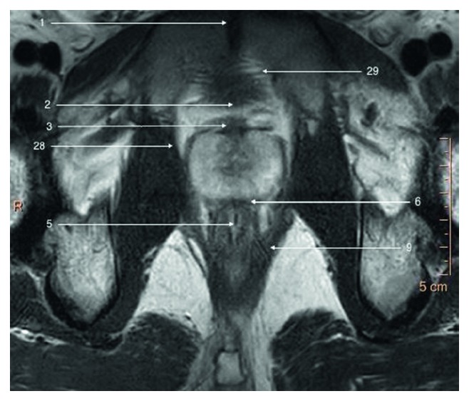
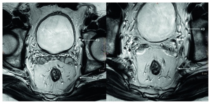
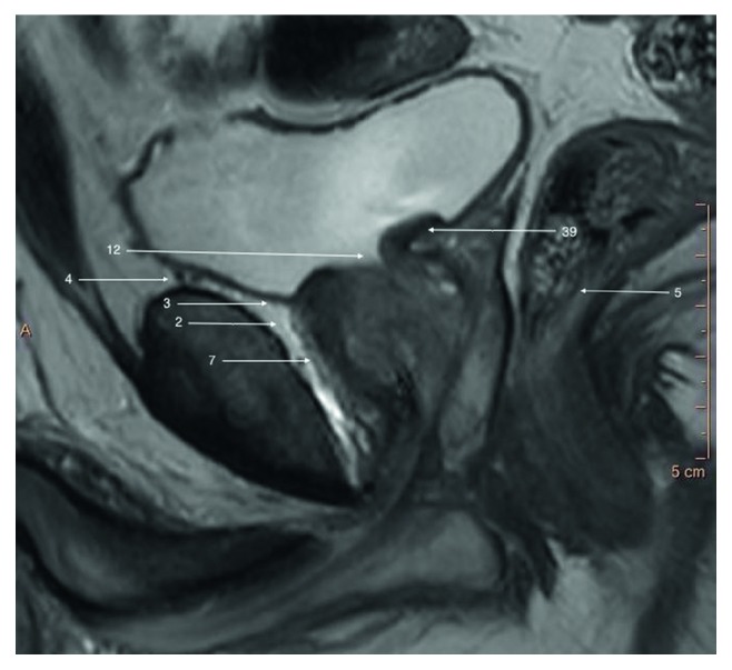
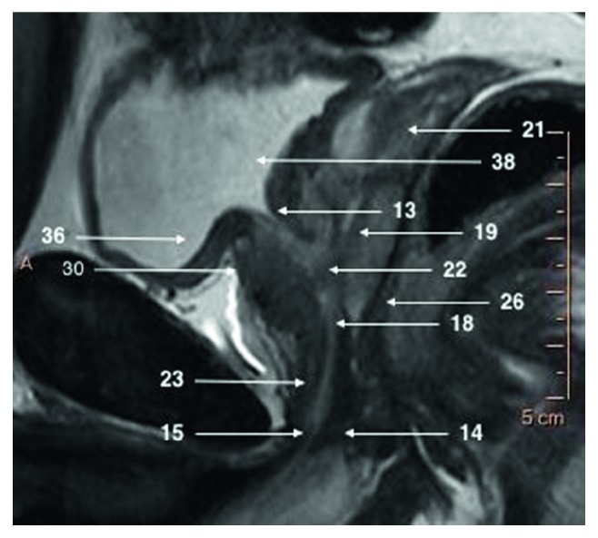
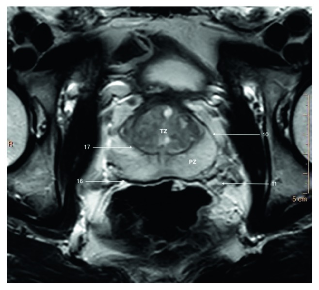
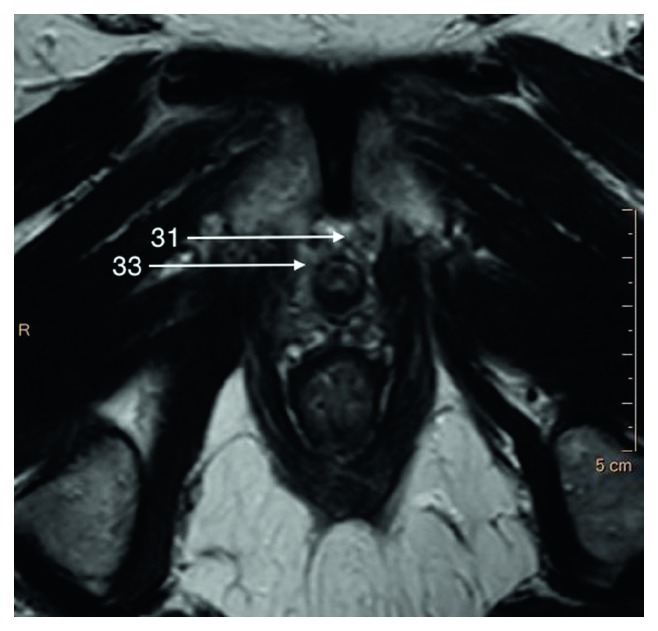
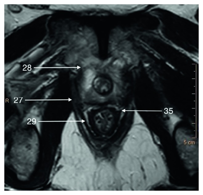
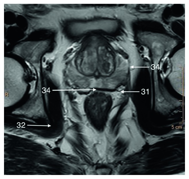
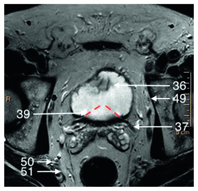
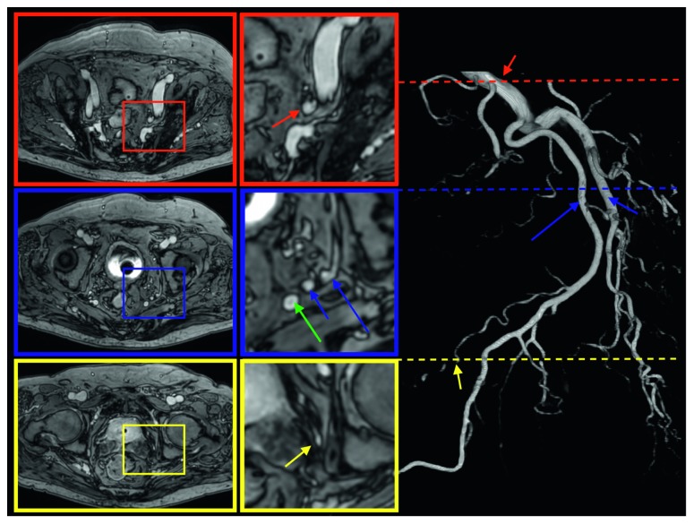
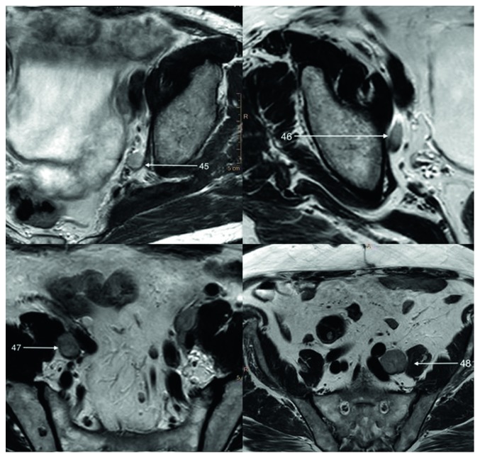
Development on new fast MRI scanners resulted in rising number of prostate examinations. High-spatial resolution of MRI examinations performed on 3T scanners allows recognition of very fine anatomical structures previously not demarcated on performed scans. We present current status of MR imaging in the context of recognition of most important anatomical structures.

摘要

新型快速磁共振成像(MRI)扫描仪的研发使得前列腺检查的数量不断增加。在3T扫描仪上进行的MRI检查具有高空间分辨率,能够识别以前在扫描中未界定的非常精细的解剖结构。我们在识别最重要解剖结构的背景下介绍了MR成像的现状。

https://cdn.ncbi.nlm.nih.gov/pmc/blobs/9542/6558623/c46a816d06b9/AMED2019-3040859.011.jpg
https://cdn.ncbi.nlm.nih.gov/pmc/blobs/9542/6558623/a80856f4bced/AMED2019-3040859.001.jpg
https://cdn.ncbi.nlm.nih.gov/pmc/blobs/9542/6558623/8c6d1bd9268a/AMED2019-3040859.002.jpg
https://cdn.ncbi.nlm.nih.gov/pmc/blobs/9542/6558623/4b09055a52a0/AMED2019-3040859.003.jpg
https://cdn.ncbi.nlm.nih.gov/pmc/blobs/9542/6558623/3c1ab44d1d94/AMED2019-3040859.004.jpg
https://cdn.ncbi.nlm.nih.gov/pmc/blobs/9542/6558623/8f5c60bfca72/AMED2019-3040859.005.jpg
https://cdn.ncbi.nlm.nih.gov/pmc/blobs/9542/6558623/4e8dae3a7460/AMED2019-3040859.006.jpg
https://cdn.ncbi.nlm.nih.gov/pmc/blobs/9542/6558623/eefd842e82a8/AMED2019-3040859.007.jpg
https://cdn.ncbi.nlm.nih.gov/pmc/blobs/9542/6558623/09e81ecfa65d/AMED2019-3040859.008.jpg
https://cdn.ncbi.nlm.nih.gov/pmc/blobs/9542/6558623/a0b328c28356/AMED2019-3040859.009.jpg
https://cdn.ncbi.nlm.nih.gov/pmc/blobs/9542/6558623/50f7d6b234c1/AMED2019-3040859.010.jpg
https://cdn.ncbi.nlm.nih.gov/pmc/blobs/9542/6558623/c46a816d06b9/AMED2019-3040859.011.jpg

相似文献

1
Normal 3T MR Anatomy of the Prostate Gland and Surrounding Structures.前列腺及周围结构的正常3T磁共振解剖
Adv Med. 2019 May 28;2019:3040859. doi: 10.1155/2019/3040859. eCollection 2019.
2
7T-Guided Learning Framework for Improving the Segmentation of 3T MR Images.用于改进3T磁共振图像分割的7T引导学习框架
Med Image Comput Comput Assist Interv. 2016 Oct;9901:572-580. doi: 10.1007/978-3-319-46723-8_66. Epub 2016 Oct 2.
3
Thirty-two-channel coil 3T magnetic resonance-guided biopsies of prostate tumor suspicious regions identified on multimodality 3T magnetic resonance imaging: technique and feasibility.在多模态3T磁共振成像上识别出的前列腺肿瘤可疑区域的32通道线圈3T磁共振引导活检:技术与可行性
Invest Radiol. 2008 Oct;43(10):686-94. doi: 10.1097/RLI.0b013e31817d0506.
4
[3 Tesla MRI: successful results with higher field strengths].[3特斯拉磁共振成像:更高场强带来的成功结果]
Radiologe. 2004 Jan;44(1):31-47. doi: 10.1007/s00117-003-1000-x.
5
Initial experience of 3 tesla endorectal coil magnetic resonance imaging and 1H-spectroscopic imaging of the prostate.3特斯拉直肠内线圈磁共振成像及前列腺1H波谱成像的初步经验
Invest Radiol. 2004 Nov;39(11):671-80. doi: 10.1097/00004424-200411000-00005.
6
Apparent diffusion coefficient and sodium concentration measurements in human prostate tissue via hydrogen-1 and sodium-23 magnetic resonance imaging in a clinical setting at 3T.在 3T 临床环境下,通过氢-1 和钠-23 磁共振成像对人前列腺组织进行表观扩散系数和钠浓度测量。
Invest Radiol. 2012 Dec;47(12):677-82. doi: 10.1097/RLI.0b013e318267bc6f.
7
MR imaging of the prostate: 1.5T versus 3T.前列腺的磁共振成像:1.5T与3T对比
Magn Reson Imaging Clin N Am. 2007 Aug;15(3):433-48, viii. doi: 10.1016/j.mric.2007.06.004.
8
Reconstruction of 7T-Like Images From 3T MRI.从3T磁共振成像重建类似7T的图像。
IEEE Trans Med Imaging. 2016 Sep;35(9):2085-97. doi: 10.1109/TMI.2016.2549918. Epub 2016 Apr 1.
9
[Clinical applications of 3.0 T magnetic resonance system in the neuroradiological field].3.0T磁共振系统在神经放射学领域的临床应用
Brain Nerve. 2007 May;59(5):479-85.
10
Hierarchical Reconstruction of 7T-like Images from 3T MRI Using Multi-level CCA and Group Sparsity.使用多级典型相关分析和组稀疏性从3T磁共振成像进行7T样图像的分层重建
Med Image Comput Comput Assist Interv. 2015 Oct;9350:659-666. doi: 10.1007/978-3-319-24571-3_79. Epub 2015 Nov 20.

引用本文的文献

1
Different sodium concentrations of noncancerous and cancerous prostate tissue seen on MRI using an external coil.使用外部线圈在MRI上观察到的非癌性和癌性前列腺组织的不同钠浓度。
Radiol Adv. 2024 Sep 30;1(3):umae023. doi: 10.1093/radadv/umae023. eCollection 2024 Sep.
2
Verification of image quality improvement by deep learning reconstruction to 1.5 T MRI in T2-weighted images of the prostate gland.利用深度学习重建技术对 1.5T MRI 前列腺 T2 加权图像进行质量改善的验证。
Radiol Phys Technol. 2024 Sep;17(3):756-764. doi: 10.1007/s12194-024-00819-5. Epub 2024 Jun 8.
3
Acquisition Parameters Influence Diffusion Metrics Effectiveness in Probing Prostate Tumor and Age-Related Microstructure.

本文引用的文献

1
Synopsis of the PI-RADS v2 Guidelines for Multiparametric Prostate Magnetic Resonance Imaging and Recommendations for Use.多参数前列腺磁共振成像PI-RADS v2指南概要及使用建议
Eur Urol. 2016 Jan;69(1):41-9. doi: 10.1016/j.eururo.2015.08.038. Epub 2015 Sep 8.
2
[The prostate gland: a crossroad between the urinary and the seminal tracts].
Prog Urol. 2012 Jun;22 Suppl 1:S2-6. doi: 10.1016/S1166-7087(12)70028-3.
3
The McNeal prostate: a review.麦克尼尔前列腺:综述。
采集参数对扩散指标探测前列腺肿瘤及与年龄相关微观结构的有效性有影响。
J Pers Med. 2023 May 20;13(5):860. doi: 10.3390/jpm13050860.
4
MRI Anatomical Investigation of Rabbit Bulbourethral Glands.兔尿道球腺的MRI解剖学研究
Animals (Basel). 2023 Apr 30;13(9):1519. doi: 10.3390/ani13091519.
5
Anterior fibromuscular stroma-preserved endoscopic enucleation of the prostate: a precision anatomical approach.保留前列腺前纤维肌肉基质的内镜前列腺剜除术:一种精准的解剖方法。
World J Urol. 2023 Aug;41(8):2127-2132. doi: 10.1007/s00345-022-04270-2. Epub 2022 Dec 30.
6
Morphometric Analysis of Prostate Zonal Anatomy After Transurethral Resection of Prostate and Holmium Laser Enucleation of Prostate Using Magnetic Resonance Imaging: A Pilot Study.经尿道前列腺切除术和钬激光前列腺剜除术后前列腺分区解剖结构的磁共振成像形态学分析:一项初步研究
Turk J Urol. 2022 May;48(3):201-208. doi: 10.5152/tud.2022.21326.
7
Imaging of clinically silent rectoprostatic hematoma in MRI guided in-bore prostate biopsy.MRI 引导经直肠前列腺穿刺活检中临床无症状直肠前列腺血肿的影像学表现。
Sci Rep. 2022 Feb 3;12(1):1840. doi: 10.1038/s41598-022-05909-1.
8
Incidental findings in and around the prostate on prostate MRI: a pictorial review.前列腺MRI检查中前列腺及其周围的偶然发现:图谱综述
Insights Imaging. 2021 Mar 18;12(1):37. doi: 10.1186/s13244-021-00979-7.
Urology. 2011 Dec;78(6):1224-8. doi: 10.1016/j.urology.2011.07.1395. Epub 2011 Sep 9.
4
Overview of prostate anatomy, histology, and pathology.前列腺解剖、组织学和病理学概述。
Endocrinol Metab Clin North Am. 2011 Sep;40(3):565-75, viii-ix. doi: 10.1016/j.ecl.2011.05.012.
5
Mapping pelvic lymph nodes: guidelines for delineation in intensity-modulated radiotherapy.盆腔淋巴结勾画:调强放射治疗中勾画指南
Int J Radiat Oncol Biol Phys. 2005 Dec 1;63(5):1604-12. doi: 10.1016/j.ijrobp.2005.05.062. Epub 2005 Sep 29.
6
Functional anatomy of the prostate: implications for treatment planning.前列腺的功能解剖学:对治疗规划的启示
Int J Radiat Oncol Biol Phys. 2005 Oct 1;63(2):479-91. doi: 10.1016/j.ijrobp.2005.02.036.