• 文献检索
  • 文档翻译
  • 深度研究
  • 学术资讯
  • Suppr Zotero 插件Zotero 插件
  • 邀请有礼
  • 套餐&价格
  • 历史记录
应用&插件
Suppr Zotero 插件Zotero 插件浏览器插件Mac 客户端Windows 客户端微信小程序
定价
高级版会员购买积分包购买API积分包
服务
文献检索文档翻译深度研究API 文档MCP 服务
关于我们
关于 Suppr公司介绍联系我们用户协议隐私条款
关注我们

Suppr 超能文献

核心技术专利:CN118964589B侵权必究
粤ICP备2023148730 号-1Suppr @ 2026

文献检索

告别复杂PubMed语法,用中文像聊天一样搜索,搜遍4000万医学文献。AI智能推荐,让科研检索更轻松。

立即免费搜索

文件翻译

保留排版,准确专业,支持PDF/Word/PPT等文件格式,支持 12+语言互译。

免费翻译文档

深度研究

AI帮你快速写综述,25分钟生成高质量综述,智能提取关键信息,辅助科研写作。

立即免费体验

动态对比增强超声评估大鼠肝癌模型组织氧状态的诊断价值。

The diagnositic value of dynamic contrast-enhanced ultrasound for evaluation of tissue oxygen status in rat hepatoma model.

机构信息

Department of ultrasound, The First Medical Centre, Chinese PLA General Hospital, No. 28 of Fuxing Road, Haidian District, Beijing, 100853, China.

出版信息

BMC Gastroenterol. 2024 Nov 23;24(1):424. doi: 10.1186/s12876-024-03523-1.

DOI:10.1186/s12876-024-03523-1
PMID:39578733
原文链接:https://pmc.ncbi.nlm.nih.gov/articles/PMC11585086/
Abstract

BACKGROUND

Hypoxia is a characteristic of solid tumors, but whether significant hypoxia exists in the hepatocellular carcinoma remains unclear. This animal study aims to explore the value of dynamic contrast-enhanced ultrasound (CEUS) quantitative parameters to evaluate the oxygen status in two rat hepatoma models.

MATERIALS AND METHODS

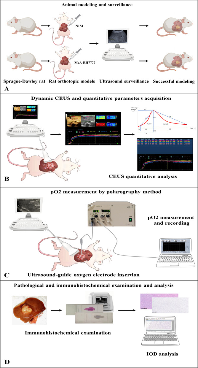
N1S1 and McA-RH7777 S-D rat orthotopic hepatoma models were established. Once the tumors reached a diameter of 10-15 mm, CEUS and oxygen partial pressure (pO2) polarography were performed. Immunohistochemical staining for HIF-1α and pimonidazole was conducted after euthanizing the rats. Correlation between quantitative CEUS parameters, pO and the immunohistochemical integrated optical density (IOD) was analyzed to assess the predictive ability of CEUS quantitative parameters for the tissue oxygen environment.

RESULT

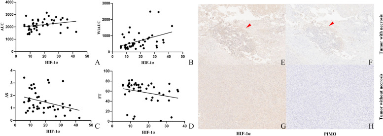
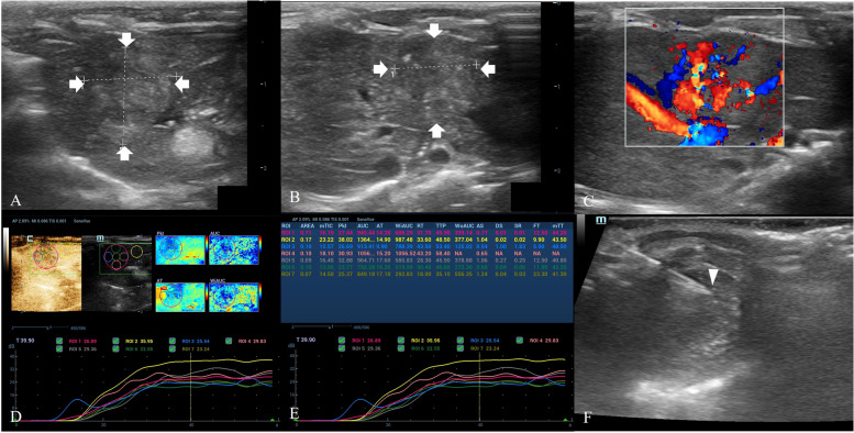
Eleven N1S1 models and ten McA-RH7777 models were established successfully. There was no significant difference in pO (35.5 mmHg vs 32.2 mmHg, P = 0.917), IOD of HIF-1α (13.4 vs 20.0, P = 0.159) and pimonidazole (0.70 vs 1.30, P = 0.926) between the tumor and the peritumoral liver tissue. The pO values were correlated with CEUS quantitative parameters including mean time-intensity curves (mTIC) (P = 0.003), peak intensity (PKI) (P = 0.010), area under the curve (AUC) (P = 0.009), area under the wash-in curve (WiAUC) (P = 0.006), and arrival time (AT) (P = 0.033). The IOD of HIF-1α correlated with AUC (P = 0.022), WiAUC (P = 0.009), ascending slope (AS) (P = 0.044), and falling time (FT) (P = 0.009). Multiple linear regression indicated that the "short AT" was an independent protective factor for hypoxia (β = -2.347, 95% CI: -4.948, -0.394, P = 0.022), and CEUS had the ability to predict the tumor pO (P = 0.003).

CONCLUSION

No evidence of significant hypoxia was identified in two rat orthotopic hepatoma models. Quantitative CEUS parameters correlated with the oxygen status of the tumor, which could be utilized to predict the tumor tissue pO.

摘要

背景

缺氧是实体瘤的一个特征,但肝癌中是否存在显著缺氧仍不清楚。本动物研究旨在探讨动态对比增强超声(CEUS)定量参数评估两种大鼠肝癌模型氧状态的价值。

材料和方法

建立 N1S1 和 McA-RH7777 S-D 大鼠原位肝癌模型。一旦肿瘤直径达到 10-15mm,进行 CEUS 和氧分压(pO2)极谱法检查。处死大鼠后进行 HIF-1α 和 pimonidazole 的免疫组织化学染色。分析定量 CEUS 参数、pO2 和免疫组化积分光密度(IOD)之间的相关性,以评估 CEUS 定量参数对组织氧环境的预测能力。

结果

成功建立了 11 个 N1S1 模型和 10 个 McA-RH7777 模型。肿瘤与肿瘤周围肝组织之间的 pO2 值(35.5mmHg 与 32.2mmHg,P=0.917)、HIF-1α 的 IOD(13.4 与 20.0,P=0.159)和 pimonidazole(0.70 与 1.30,P=0.926)无显著差异。pO2 值与 CEUS 定量参数相关,包括平均时间强度曲线(mTIC)(P=0.003)、峰值强度(PKI)(P=0.010)、曲线下面积(AUC)(P=0.009)、曲线上升下面积(WiAUC)(P=0.006)和到达时间(AT)(P=0.033)。HIF-1α 的 IOD 与 AUC(P=0.022)、WiAUC(P=0.009)、上升斜率(AS)(P=0.044)和下降时间(FT)(P=0.009)相关。多元线性回归表明,“短 AT”是缺氧的独立保护因素(β=-2.347,95%CI:-4.948,-0.394,P=0.022),CEUS 具有预测肿瘤 pO2 的能力(P=0.003)。

结论

在两种大鼠原位肝癌模型中未发现明显缺氧。定量 CEUS 参数与肿瘤的氧状态相关,可用于预测肿瘤组织的 pO2。

https://cdn.ncbi.nlm.nih.gov/pmc/blobs/4c5a/11585086/1abfafac04ee/12876_2024_3523_Fig4_HTML.jpg
https://cdn.ncbi.nlm.nih.gov/pmc/blobs/4c5a/11585086/7c18f2c85f74/12876_2024_3523_Fig1_HTML.jpg
https://cdn.ncbi.nlm.nih.gov/pmc/blobs/4c5a/11585086/50c7c70af65d/12876_2024_3523_Fig2_HTML.jpg
https://cdn.ncbi.nlm.nih.gov/pmc/blobs/4c5a/11585086/7538d371a977/12876_2024_3523_Fig3_HTML.jpg
https://cdn.ncbi.nlm.nih.gov/pmc/blobs/4c5a/11585086/1abfafac04ee/12876_2024_3523_Fig4_HTML.jpg

相似文献

1
The diagnositic value of dynamic contrast-enhanced ultrasound for evaluation of tissue oxygen status in rat hepatoma model.动态对比增强超声评估大鼠肝癌模型组织氧状态的诊断价值。
BMC Gastroenterol. 2024 Nov 23;24(1):424. doi: 10.1186/s12876-024-03523-1.
2
Comparison of dynamic contrast-enhanced magnetic resonance imaging and contrast-enhanced ultrasound for evaluation of the effects of sorafenib in a rat model of hepatocellular carcinoma.动态对比增强磁共振成像与超声造影在评价索拉非尼对大鼠肝细胞癌模型疗效中的比较
Magn Reson Imaging. 2019 Apr;57:156-164. doi: 10.1016/j.mri.2018.11.012. Epub 2018 Nov 19.
3
Therapeutic response monitoring after targeted therapy in an orthotopic rat model of hepatocellular carcinoma using contrast-enhanced ultrasound: Focusing on inter-scanner, and inter-operator reproducibility.应用超声造影监测肝细胞癌大鼠模型中靶向治疗的疗效:重点探讨扫描仪间和操作者间的可重复性。
PLoS One. 2020 Dec 23;15(12):e0244304. doi: 10.1371/journal.pone.0244304. eCollection 2020.
4
Potential application of dynamic contrast enhanced ultrasound in predicting microvascular invasion of hepatocellular carcinoma.动态对比增强超声在预测肝细胞癌微血管侵犯中的应用潜力。
Clin Hemorheol Microcirc. 2021;77(4):461-469. doi: 10.3233/CH-201085.
5
Modified Rat Hepatocellular Carcinoma Models Overexpressing Vascular Endothelial Growth Factor.过表达血管内皮生长因子的改良大鼠肝细胞癌模型
J Vasc Interv Radiol. 2018 Nov;29(11):1604-1612. doi: 10.1016/j.jvir.2018.07.005. Epub 2018 Oct 5.
6
Hepatic Artery Embolization Enhances Expression of Programmed Cell Death 1 Ligand 1 in an Orthotopic Rat Hepatocellular Carcinoma Model: In Vivo and in Vitro Experimentation.肝动脉栓塞增强原位大鼠肝细胞癌模型中程序性细胞死亡蛋白1配体1的表达:体内和体外实验
J Vasc Interv Radiol. 2020 Sep;31(9):1475-1482.e2. doi: 10.1016/j.jvir.2020.03.023. Epub 2020 Aug 14.
7
Quantitative analysis of hepatic hypoxia-inducible factor-1alpha and its abnormal gene expression during the formation of hepatocellular carcinoma.定量分析肝癌形成过程中肝缺氧诱导因子-1α及其异常基因表达。
Hepatobiliary Pancreat Dis Int. 2009 Aug;8(4):407-13.
8
Comparison of tumor vascularity and hemodynamics in three rat hepatoma models.三种大鼠肝癌模型中肿瘤血管生成与血流动力学的比较。
Abdom Radiol (NY). 2016 Feb;41(2):257-64. doi: 10.1007/s00261-015-0591-9.
9
Effects of hypoxia-inducible factor-1α silencing on the proliferation of CBRH-7919 hepatoma cells.缺氧诱导因子-1α 沉默对 CBRH-7919 肝癌细胞增殖的影响。
World J Gastroenterol. 2013 Mar 21;19(11):1749-59. doi: 10.3748/wjg.v19.i11.1749.
10
Quantitative perfusion analysis of hepatocellular carcinoma using dynamic contrast enhanced ultrasound (CEUS) to determine tumor microvascularization.使用动态对比增强超声(CEUS)定量分析肝细胞癌以确定肿瘤微血管化。
Clin Hemorheol Microcirc. 2019;73(1):95-104. doi: 10.3233/CH-199221.

本文引用的文献

1
Simultaneous photoacoustic and ultrasound imaging: A review.同时光声和超声成像:综述。
Ultrasonics. 2024 Apr;139:107277. doi: 10.1016/j.ultras.2024.107277. Epub 2024 Feb 28.
2
Cabozantinib plus atezolizumab versus sorafenib for advanced hepatocellular carcinoma (COSMIC-312): final results of a randomised phase 3 study.卡博替尼联合阿替利珠单抗对比索拉非尼治疗晚期肝细胞癌(COSMIC-312):一项随机3期研究的最终结果
Lancet Gastroenterol Hepatol. 2024 Apr;9(4):310-322. doi: 10.1016/S2468-1253(23)00454-5. Epub 2024 Feb 13.
3
CALGB 80802 (Alliance): Impact of Sorafenib with and without Doxorubicin on Hepatitis C Infection in Patients with Advanced Hepatocellular Carcinoma.
CALGB 80802(Alliance):索拉非尼联合或不联合多柔比星对晚期肝细胞癌合并丙型肝炎感染患者的影响。
Cancer Res Commun. 2024 Mar 7;4(3):682-690. doi: 10.1158/2767-9764.CRC-22-0516.
4
Current status of optoacoustic breast imaging and future trends in clinical application: is it ready for prime time?光声乳腺成像的现状及临床应用的未来趋势:是否已准备好投入使用?
Eur Radiol. 2024 Sep;34(9):6092-6107. doi: 10.1007/s00330-024-10600-2. Epub 2024 Feb 3.
5
Cancer statistics, 2024.2024年癌症统计数据。
CA Cancer J Clin. 2024 Jan-Feb;74(1):12-49. doi: 10.3322/caac.21820. Epub 2024 Jan 17.
6
Pancreatic Ductal Adenocarcinoma: The Characteristics of Contrast-Enhanced Ultrasound Are Correlated with the Hypoxic Microenvironment.胰腺导管腺癌:超声造影特征与缺氧微环境相关
Diagnostics (Basel). 2023 Oct 20;13(20):3270. doi: 10.3390/diagnostics13203270.
7
Photoacoustic imaging for investigating tumor hypoxia: a strategic assessment.光声成像技术用于研究肿瘤乏氧:策略评估
Theranostics. 2023 May 29;13(10):3346-3367. doi: 10.7150/thno.84253. eCollection 2023.
8
1D-model of the human liver circulatory system.人体肝脏循环系统的 1D 模型。
Comput Methods Programs Biomed. 2023 Aug;238:107612. doi: 10.1016/j.cmpb.2023.107612. Epub 2023 May 16.
9
Evaluation of Hypoxia in Hepatocellular Carcinoma Using Quantitative MRI: Significances, Challenges, and Advances.利用定量 MRI 评估肝细胞癌的缺氧:意义、挑战和进展。
J Magn Reson Imaging. 2023 Jul;58(1):12-25. doi: 10.1002/jmri.28694. Epub 2023 Mar 27.
10
Severe hypoxia is a typical characteristic of human hepatocellular carcinoma: Scientific fact or fallacy?严重缺氧是人类肝细胞癌的典型特征:科学事实还是谬误?
J Hepatol. 2022 Apr;76(4):975-980. doi: 10.1016/j.jhep.2021.12.028. Epub 2022 Jan 3.