Oukili Houssine, Benlghazi Abdelhamid, Benmoussa Meryem, Outaghyame Oussama, Elhassani Moulay Mehdi, Kouach Jaouad
Department of Gynecology-Obstetrics, Mohammed V Military Teaching Hospital, Faculty of Medicine and Pharmacy of Rabat, University Mohammed V, Rabat, Morocco.
Department of Gynecology-Obstetrics, Mohammed V Military Teaching Hospital, Faculty of Medicine and Pharmacy of Rabat, University Mohammed V, Rabat, Morocco.
Int J Surg Case Rep. 2024 Dec;125:110620. doi: 10.1016/j.ijscr.2024.110620. Epub 2024 Nov 16.
Uterine arteriovenous malformations are a rare but potentially life-threatening condition. They may be congenital or acquired and should be suspected in cases of severe or persistent uterine hemorrhage.
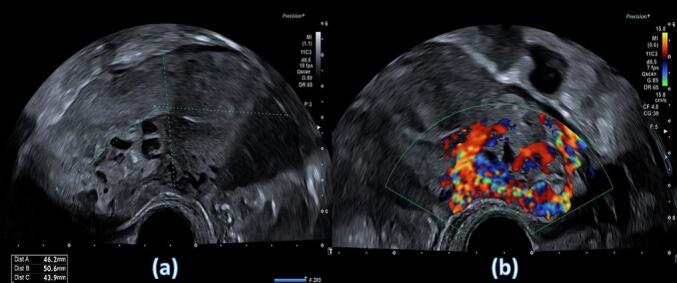
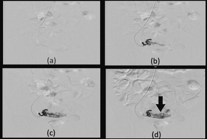
We present the clinical case of a 32-year-old woman who suffered from bleeding after a spontaneous miscarriage. Uterine arteriovenous malformation with trophoblastic retention was suspected on ultrasound and Doppler examination. Magnetic resonance imaging confirmed the diagnosis, leading to embolization of both uterine arteries, followed by operative hysteroscopy to remove the retained tissue, with a favorable outcome.
Uterine arteriovenous malformation is rare, with fewer than 100 cases reported in the literature (1). It is a potentially fatal condition due to the heavy bleeding that patients may experience. Color Doppler ultrasound (US) is a non-invasive method for initially diagnosing this rare condition, which can be confirmed by diagnostic angiography. A conservative approach or embolization is the preferred treatment to avoid hysterectomy in patients of childbearing age.
This case report emphasizes the use of ultrasound and MRI to diagnose a uterine AVM in a patient of childbearing age who presented with post-partum retention of products. It also showcases our experience with embolization in this patient, which allowed her to preserve her fertility.
子宫动静脉畸形是一种罕见但可能危及生命的疾病。它们可能是先天性的或后天获得的,在严重或持续性子宫出血的病例中应怀疑此病。
我们呈现了一名32岁女性的临床病例,该女性在自然流产后出现出血。超声和多普勒检查怀疑为伴有滋养细胞残留的子宫动静脉畸形。磁共振成像确诊了该疾病,随后对双侧子宫动脉进行栓塞,接着进行手术宫腔镜检查以清除残留组织,结果良好。
子宫动静脉畸形较为罕见,文献报道的病例少于100例(1)。由于患者可能出现大量出血,这是一种潜在致命的疾病。彩色多普勒超声(US)是初步诊断这种罕见疾病的非侵入性方法,可通过诊断性血管造影加以确诊。对于育龄期患者,保守治疗或栓塞是避免子宫切除术的首选治疗方法。
本病例报告强调了在一名产后有产物残留的育龄期患者中使用超声和MRI诊断子宫动静脉畸形。它还展示了我们对该患者进行栓塞治疗的经验,使她能够保留生育能力。