Shimoda Marine Research Center, University of Tsukuba, 5-10-1, Shimoda 415-0025, Shizuoka, Japan.
Faculty of Life Science and Biotechnology, Fukuyama University, Fukuyama 729-0292, Hiroshima, Japan.
Cells. 2024 Nov 11;13(22):1863. doi: 10.3390/cells13221863.
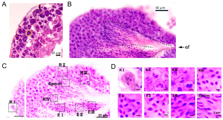
Animals show diverse processes of gametogenesis in the evolutionary pathway. Here, we characterized the spermatogenic cells in the testis of the marine invertebrate sperm differentiate in a non-cystic type of testis, comprising many follicles with various sizes and stages of spermatogenic cells. In the space among follicles, we observed free cells that were recognized by antibody against Müllerian inhibiting substance, a marker for vertebrate Sertoli cells. We further categorized the spermatogenic cells into four round stages (RI to RIV) and three elongated stages (EI to EIII) by morphological and immunohistochemical criteria. An antibody against a vertebrate Vasa homolog recognized a few large spermatogonium-like cells (RI) near the basal wall of a follicle. Consistent with the period of meiosis, a synaptonemal complex protein SYCP3 was recognized from early spermatocytes (RII) to early spermatids (E1). Acetylated tubulins were detected in spermatids before flagellar elongation at the RIV stage and became distributed along the flagella. Electron microscopy showed that the free cells outside the testicular follicle possessed a characteristic of vertebrate Sertoli cells. These results would provide a basis for basic and comparative studies on the mechanism of spermatogenesis.
动物在进化过程中表现出多样化的配子发生过程。在这里,我们描述了海洋无脊椎动物 精子发生的精巢中的生精细胞。在精巢中,生精细胞分化为非囊泡型,包含许多具有不同大小和生精细胞阶段的滤泡。在滤泡之间的空间中,我们观察到了自由细胞,这些细胞被抗 Müllerian 抑制物质的抗体识别,Müllerian 抑制物质是脊椎动物 Sertoli 细胞的标志物。我们进一步根据形态学和免疫组织化学标准将生精细胞分为四个圆形阶段(RI 到 RIV)和三个伸长阶段(EI 到 EIII)。一种针对脊椎动物 Vasa 同源物的抗体识别出靠近滤泡基底壁的几个大精原细胞样细胞(RI)。与减数分裂时期一致,从早期精母细胞(RII)到早期精子(E1)都能识别出联会复合体蛋白 SYCP3。在 RIV 阶段鞭毛伸长之前,乙酰化微管在精子中被检测到,并沿鞭毛分布。电子显微镜显示,滤泡外的自由细胞具有脊椎动物 Sertoli 细胞的特征。这些结果将为基础和比较研究精子发生机制提供依据。