Park Hee-Jung, Nam Myeong-Hyun, Park Ji-Hoon, Lee Ji-Min, Hong Hye-Sun, Kim Tae-Woo, Lee In-Ho, Shin Chang-Ho, Lee Soo-Hong, Seo Young-Kwon
Department of Biomedical Engineering, Dongguk University, Goyang-si 10326, Republic of Korea.
Department of AI Convergence Biomedical Engineering, Dongguk University, Goyang-si 10326, Republic of Korea.
Biomedicines. 2024 Oct 28;12(11):2475. doi: 10.3390/biomedicines12112475.
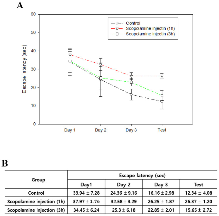
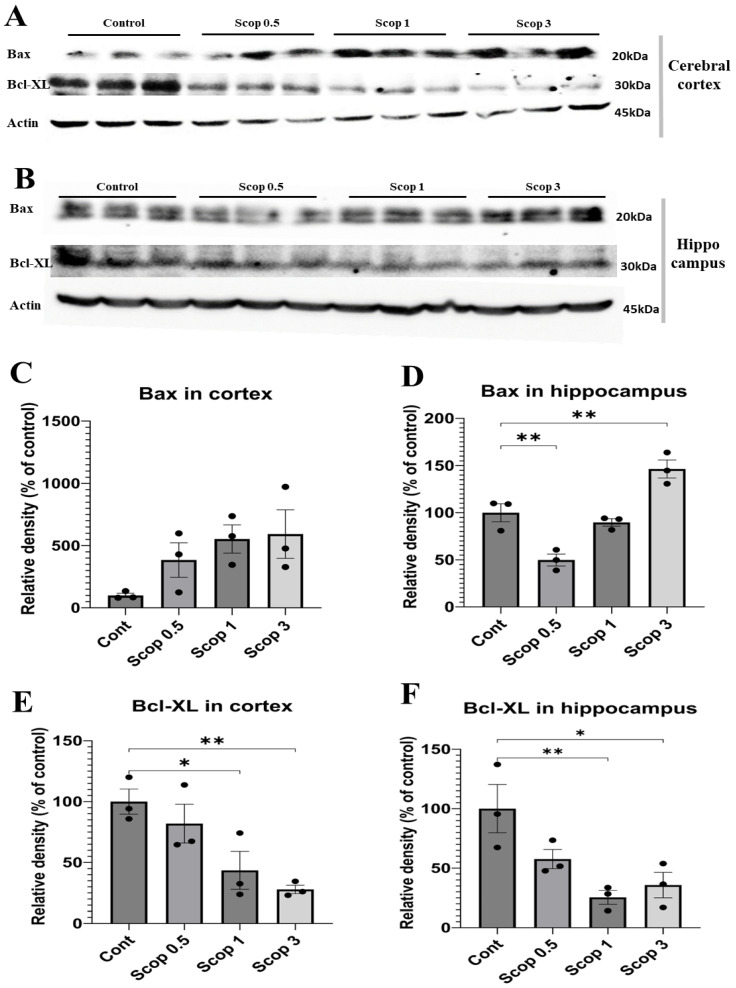
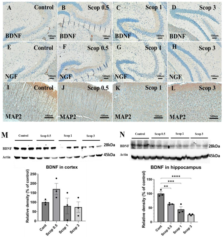
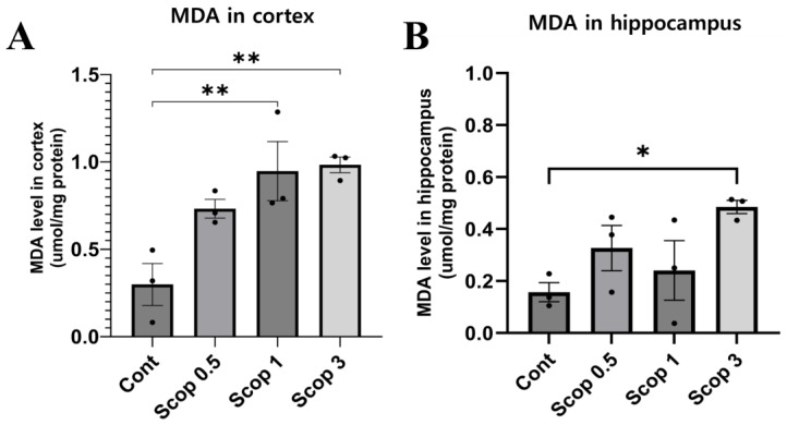
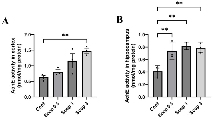
Until now, many researchers have conducted evaluations on hippocampi for analyses of cognitive dysfunction models using scopolamine. However, depending on the purposes of these analyses, there are differences in the experimental results for the hippocampi and cortexes. Therefore, this study intends to compare various analyses of cognitive dysfunction after scopolamine administration with each other in hippocampi and cortexes. Scopolamine was administered at three dosages in mice: 0.5, 1, and 3 mg/kg. And this study evaluates the differences in cognitive function and the expression of malondialdehyde (MDA), acetylcholinesterase (AChE), and brain-derived neurotrophic factor (BDNF) in mice's hippocampi and cortexes based on scopolamine dosages. The Morris water maze test was conducted between 1 and 3 h after scopolamine injection to assess its duration. A significant decrease in behavioral ability was evaluated at 1 h, and we observed a similar recovery to the normal group at 3 h. And the Morris water maze escape latency showed differences depending on scopolamine concentration. While the escape waiting time in the control group and scop 0.5 administration group remained similar to that seen before administration, the administration of scop 1 and 3 increased it. In the experimental group administered scop 1 and 3, cerebral MDA levels in the cerebral cortex significantly increased. In the hippocampus, the MDA level in the scopolamine-administered groups slightly increased compared to the cortex. A Western blotting assay shows that Bax and Bcl-xl showed a tendency to increase or decrease depending on the concentration, but BDNF increased in scop 0.5, and scop 1 and 3 did not show a significant decrease compared to the control at the cerebral cortex. In the hippocampus, BDNF showed a concentration-dependent decrease in expression. This study's findings indicate that chemical analyses for MDA and AChE can be performed in the cerebral cortex, while the hippocampus is better suited for protein analysis of apoptosis and BDNF.
到目前为止,许多研究人员已对海马体进行评估,以分析使用东莨菪碱的认知功能障碍模型。然而,根据这些分析的目的,海马体和皮质的实验结果存在差异。因此,本研究旨在比较东莨菪碱给药后海马体和皮质中认知功能障碍的各种分析。东莨菪碱以三种剂量给予小鼠:0.5、1和3mg/kg。本研究基于东莨菪碱剂量评估小鼠海马体和皮质中认知功能的差异以及丙二醛(MDA)、乙酰胆碱酯酶(AChE)和脑源性神经营养因子(BDNF)的表达。在注射东莨菪碱后1至3小时进行莫里斯水迷宫试验以评估其持续时间。在1小时时评估行为能力显著下降,并且我们在3小时时观察到与正常组相似的恢复。并且莫里斯水迷宫逃避潜伏期根据东莨菪碱浓度显示出差异。虽然对照组和东莨菪碱0.5给药组的逃避等待时间与给药前相似,但东莨菪碱1和3给药组的逃避等待时间增加。在给予东莨菪碱1和3的实验组中,大脑皮质中的脑MDA水平显著增加。在海马体中,与皮质相比,东莨菪碱给药组中的MDA水平略有增加。蛋白质印迹分析表明,Bax和Bcl-xl显示出根据浓度增加或减少的趋势,但在大脑皮质中,BDNF在东莨菪碱0.5给药组中增加,东莨菪碱1和3给药组与对照组相比没有显著下降。在海马体中,BDNF表达呈浓度依赖性下降。本研究结果表明,可以在大脑皮质中进行MDA和AChE的化学分析,而海马体更适合进行细胞凋亡和BDNF的蛋白质分析。