• 文献检索
  • 文档翻译
  • 深度研究
  • 学术资讯
  • Suppr Zotero 插件Zotero 插件
  • 邀请有礼
  • 套餐&价格
  • 历史记录
应用&插件
Suppr Zotero 插件Zotero 插件浏览器插件Mac 客户端Windows 客户端微信小程序
定价
高级版会员购买积分包购买API积分包
服务
文献检索文档翻译深度研究API 文档MCP 服务
关于我们
关于 Suppr公司介绍联系我们用户协议隐私条款
关注我们

Suppr 超能文献

核心技术专利:CN118964589B侵权必究
粤ICP备2023148730 号-1Suppr @ 2026

文献检索

告别复杂PubMed语法,用中文像聊天一样搜索,搜遍4000万医学文献。AI智能推荐,让科研检索更轻松。

立即免费搜索

文件翻译

保留排版,准确专业,支持PDF/Word/PPT等文件格式,支持 12+语言互译。

免费翻译文档

深度研究

AI帮你快速写综述,25分钟生成高质量综述,智能提取关键信息,辅助科研写作。

立即免费体验

基于前列腺周围脂肪磁共振成像的放射组学列线图预测根治性前列腺切除术后非转移性前列腺癌患者的生化无复发生存。

Periprostatic fat magnetic resonance imaging based radiomics nomogram for predicting biochemical recurrence-free survival in patients with non-metastatic prostate cancer after radical prostatectomy.

机构信息

Department of Urology, Urology Research Institute, the First Affiliated Hospital, Fujian Medical University, Fuzhou, 350005, China.

Department of Urology, Binhai Campus of the First Affiliated Hospital, National Region Medical centre, Fujian Medical University, Fuzhou, 350212, China.

出版信息

BMC Cancer. 2024 Nov 27;24(1):1459. doi: 10.1186/s12885-024-13207-4.

DOI:10.1186/s12885-024-13207-4
PMID:39604917
原文链接:https://pmc.ncbi.nlm.nih.gov/articles/PMC11600801/
Abstract

OBJECTIVE

To build and validate a periprostatic fat magnetic resonance imaging (MRI) based radiomics nomogram for prediction of biochemical recurrence-free survival (bRFS) of patients with non-metastatic prostate cancer (PCa) receiving radical prostatectomy (RP).

METHODS

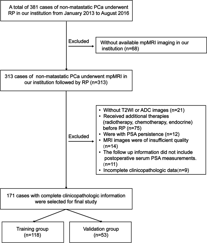
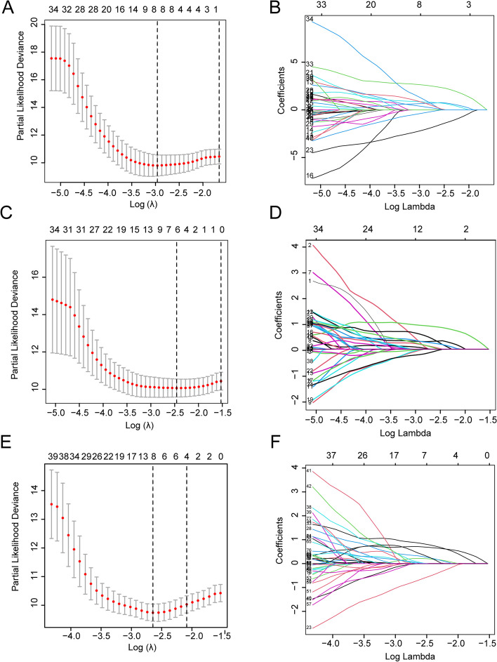
A retrospective study was conducted on 356 patients with non-metastatic PCa who underwent preoperative mpMRI followed by RP treatment at our institution. Radiomic features were extracted from both intratumoral region and the periprostatic fat region, which were segmented on images obtained through T2-weighted imaging (T2WI) and apparent-diffusion coefficient (ADC) imaging. Three radiomics models were developed by applying the Least absolute shrinkage and selection operator (LASSO) Cox regression, followed by Cox risk regression to construct a combined radiomics-clinical model by integrating the optimal radiomics score and clinicopathological risk factors to draw a nomogram. The predictive performance was evaluated using receiver operating characteristic (ROC) curves, Kaplan-Meier analysis and calibration curves.

RESULTS

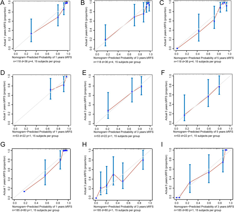
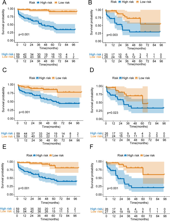
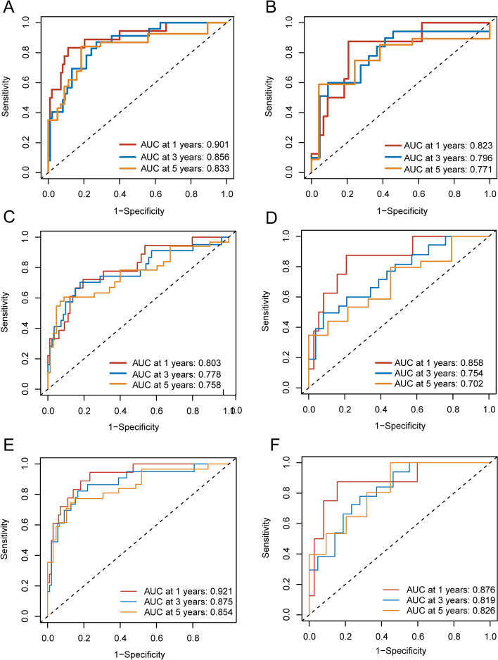
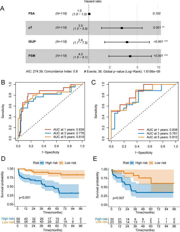
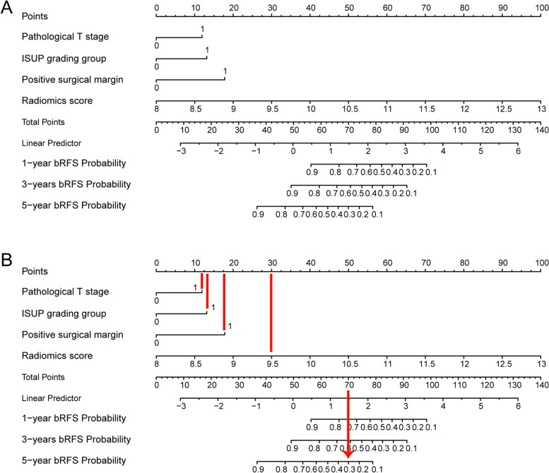
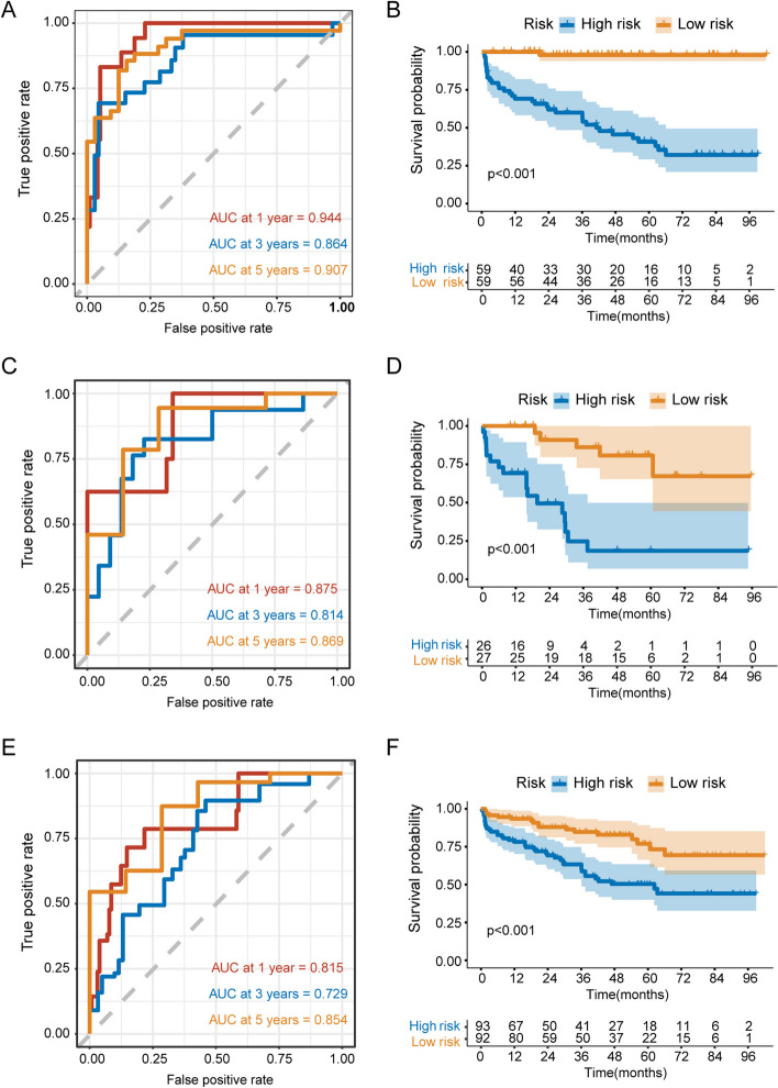
One hundred and twenty-one patients (33.98%) experienced biochemical recurrence. ROC analyses showed that the Area Under the Curve (AUC) of the periprostatic fat-intratumoral radiomics model demonstrated the highest AUC at 0.921 (95%CI, 0.857-0.981), 0.875 (95%CI, 0.763-0.950), 0.854 (95%CI, 0.706-0.923) for 1-year, 3-years and 5-years bRFS. Multivariate Cox regression analysis revealed that Pathological T stage, ISUP grading group and Positive surgical margin were independent prognostic factors for predicting bRFS. A radiomics-clinical nomogram based on these clinical predictors and periprostatic fat-intratumoral radiomics score was constructed. Kaplan-Meier analyses showed that radiomics-clinical nomogram was significantly related with survival of PCa (P < 0.001); and calibration curves revealed the predicted and observed survival probability of 1-year, 3-year and 5-year bRFS had high degree of consistency in the training and validation group. The radiomics-clinical nomogram showed a significant improvement than the clinical model for 1-year (AUC, 0.944; 95%CI, 0.912-0.990 vs. AUC, 0.839; 95%CI, 0.661-0.928; P = 0.009), 3-year (AUC, 0.864; 95%CI, 0.772-0.969 vs. AUC, 0.776; 95%CI, 0.602-0.872; P = 0.008), and 5-year bRFS (AUC, 0.907; 95%CI, 0.836-0.982 vs. AUC, 0.819; 95%CI, 0.687-0.915; P = 0.027).

CONCLUSIONS

This study developed and validated the radiomics-clinical nomogram for the prediction of bRFS in non-metastatic PCa patients underwent RP.

摘要

目的

建立并验证基于前列腺周围脂肪磁共振成像(MRI)的放射组学列线图,以预测接受根治性前列腺切除术(RP)治疗的非转移性前列腺癌(PCa)患者的生化无复发生存率(bRFS)。

方法

对在我院接受术前 mpMRI 检查并随后接受 RP 治疗的 356 例非转移性 PCa 患者进行回顾性研究。从 T2 加权成像(T2WI)和表观扩散系数(ADC)图像上勾画的肿瘤内区域和前列腺周围脂肪区域提取放射组学特征。应用最小绝对收缩和选择算子(LASSO)Cox 回归分析,构建三个放射组学模型,然后通过 Cox 风险回归分析,将最佳放射组学评分和临床病理危险因素相结合,构建一个联合放射组学-临床模型,绘制列线图。通过接受者操作特征(ROC)曲线、Kaplan-Meier 分析和校准曲线来评估预测性能。

结果

121 例患者(33.98%)发生生化复发。ROC 分析显示,前列腺周围脂肪-肿瘤内放射组学模型的曲线下面积(AUC)在 1 年、3 年和 5 年 bRFS 中表现最佳,分别为 0.921(95%CI,0.857-0.981)、0.875(95%CI,0.763-0.950)和 0.854(95%CI,0.706-0.923)。多变量 Cox 回归分析显示,病理 T 分期、ISUP 分级组和阳性切缘是预测 bRFS 的独立预后因素。基于这些临床预测因素和前列腺周围脂肪-肿瘤内放射组学评分构建了一个放射组学-临床列线图。Kaplan-Meier 分析显示,放射组学-临床列线图与 PCa 患者的生存情况显著相关(P<0.001);校准曲线显示,1 年、3 年和 5 年 bRFS 的预测和观察生存概率在训练组和验证组中具有高度一致性。与临床模型相比,放射组学-临床列线图在 1 年(AUC,0.944;95%CI,0.912-0.990 vs. AUC,0.839;95%CI,0.661-0.928;P=0.009)、3 年(AUC,0.864;95%CI,0.772-0.969 vs. AUC,0.776;95%CI,0.602-0.872;P=0.008)和 5 年 bRFS(AUC,0.907;95%CI,0.836-0.982 vs. AUC,0.819;95%CI,0.687-0.915;P=0.027)方面均有显著改善。

结论

本研究建立并验证了基于前列腺周围脂肪磁共振成像的放射组学-临床列线图,用于预测接受根治性前列腺切除术治疗的非转移性前列腺癌患者的生化无复发生存率。

https://cdn.ncbi.nlm.nih.gov/pmc/blobs/74e8/11600801/870cbad120f2/12885_2024_13207_Fig8_HTML.jpg
https://cdn.ncbi.nlm.nih.gov/pmc/blobs/74e8/11600801/ced9c96ace9a/12885_2024_13207_Fig1_HTML.jpg
https://cdn.ncbi.nlm.nih.gov/pmc/blobs/74e8/11600801/4237518a4424/12885_2024_13207_Fig2_HTML.jpg
https://cdn.ncbi.nlm.nih.gov/pmc/blobs/74e8/11600801/ebdb9aa8f3ea/12885_2024_13207_Fig3_HTML.jpg
https://cdn.ncbi.nlm.nih.gov/pmc/blobs/74e8/11600801/1581fb2e462c/12885_2024_13207_Fig4_HTML.jpg
https://cdn.ncbi.nlm.nih.gov/pmc/blobs/74e8/11600801/868ac7d3f7c7/12885_2024_13207_Fig5_HTML.jpg
https://cdn.ncbi.nlm.nih.gov/pmc/blobs/74e8/11600801/3536454aaa84/12885_2024_13207_Fig6_HTML.jpg
https://cdn.ncbi.nlm.nih.gov/pmc/blobs/74e8/11600801/9ef523a2ff0e/12885_2024_13207_Fig7_HTML.jpg
https://cdn.ncbi.nlm.nih.gov/pmc/blobs/74e8/11600801/870cbad120f2/12885_2024_13207_Fig8_HTML.jpg

相似文献

1
Periprostatic fat magnetic resonance imaging based radiomics nomogram for predicting biochemical recurrence-free survival in patients with non-metastatic prostate cancer after radical prostatectomy.基于前列腺周围脂肪磁共振成像的放射组学列线图预测根治性前列腺切除术后非转移性前列腺癌患者的生化无复发生存。
BMC Cancer. 2024 Nov 27;24(1):1459. doi: 10.1186/s12885-024-13207-4.
2
Magnetic resonance imaging-based radiomics nomogram for the evaluation of therapeutic responses to neoadjuvant chemohormonal therapy in high-risk non-metastatic prostate cancer.基于磁共振成像的放射组学列线图用于评估高危非转移性前列腺癌新辅助化疗内分泌治疗的疗效。
Cancer Med. 2024 Jul;13(14):e70001. doi: 10.1002/cam4.70001.
3
Development and Validation of an MRI-Based Radiomics Nomogram to Predict the Prognosis of De Novo Oligometastatic Prostate Cancer Patients.基于MRI的影像组学列线图预测初发寡转移前列腺癌患者预后的开发与验证
Cancer Med. 2024 Dec;13(24):e70481. doi: 10.1002/cam4.70481.
4
A dynamic online nomogram predicting prostate cancer short-term prognosis based on F-PSMA-1007 PET/CT of periprostatic adipose tissue: a multicenter study.一项基于前列腺周围脂肪组织的F-PSMA-1007 PET/CT预测前列腺癌短期预后的动态在线列线图:一项多中心研究。
Abdom Radiol (NY). 2024 Oct;49(10):3747-3757. doi: 10.1007/s00261-024-04421-6. Epub 2024 Jun 18.
5
A novel imaging based Nomogram for predicting post-surgical biochemical recurrence and adverse pathology of prostate cancer from pre-operative bi-parametric MRI.基于新型影像学的列线图预测前列腺癌患者术前双参数 MRI 与术后生化复发及不良病理的关系。
EBioMedicine. 2021 Jan;63:103163. doi: 10.1016/j.ebiom.2020.103163. Epub 2020 Dec 13.
6
Development and validation of [18 F]-PSMA-1007 PET-based radiomics model to predict biochemical recurrence-free survival following radical prostatectomy.基于[18F]-PSMA-1007 PET 的放射组学模型的开发和验证,用于预测根治性前列腺切除术后生化无复发生存。
Eur J Nucl Med Mol Imaging. 2024 Jul;51(9):2806-2818. doi: 10.1007/s00259-024-06734-6. Epub 2024 May 1.
7
A Radiomics nomogram for predicting bone metastasis in newly diagnosed prostate cancer patients.用于预测初诊前列腺癌患者骨转移的放射组学列线图
Eur J Radiol. 2020 Jul;128:109020. doi: 10.1016/j.ejrad.2020.109020. Epub 2020 Apr 19.
8
Ultrasound-based nomogram to predict the recurrence in papillary thyroid carcinoma using machine learning.基于超声的列线图,使用机器学习预测甲状腺乳头状癌的复发。
BMC Cancer. 2024 Jul 7;24(1):810. doi: 10.1186/s12885-024-12546-6.
9
Radiomics Based on MRI as a Biomarker to Guide Therapy by Predicting Upgrading of Prostate Cancer From Biopsy to Radical Prostatectomy.基于磁共振成像(MRI)的影像组学作为一种生物标志物,通过预测前列腺癌从活检到根治性前列腺切除术的升级来指导治疗。
J Magn Reson Imaging. 2020 Oct;52(4):1239-1248. doi: 10.1002/jmri.27138. Epub 2020 Mar 17.
10
Preoperative prediction of histopathological grading in patients with chondrosarcoma using MRI-based radiomics with semantic features.基于 MRI 影像组学和语义特征的术前预测软骨肉瘤患者的组织病理学分级。
BMC Med Imaging. 2024 Jul 11;24(1):171. doi: 10.1186/s12880-024-01330-4.

引用本文的文献

1
Radiologic Assessment of Periprostatic Fat as an Indicator of Prostate Cancer Risk on Multiparametric MRI.经多参数磁共振成像评估前列腺周围脂肪作为前列腺癌风险指标的影像学研究
Bioengineering (Basel). 2025 Jul 31;12(8):831. doi: 10.3390/bioengineering12080831.
2
CT-based radiomics model for noninvasive prediction of progression-free survival in high-grade serous ovarian carcinoma: a multicenter study incorporating preoperative and postoperative clinical factors.基于CT的影像组学模型用于高级别浆液性卵巢癌无进展生存期的无创预测:一项纳入术前和术后临床因素的多中心研究
BMC Med Imaging. 2025 Aug 8;25(1):320. doi: 10.1186/s12880-025-01865-0.
3
MRI-based radiomics for prediction of biochemical recurrence in prostate cancer: a systematic review and meta-analysis.
基于MRI的影像组学预测前列腺癌生化复发:一项系统综述和荟萃分析
Abdom Radiol (NY). 2025 Mar 27. doi: 10.1007/s00261-025-04892-1.