Facultad de Ciencias Biológicas, Pontificia Universidad Católica de Chile, Santiago, Chile.
Institute for Biological and Medical Engineering, Schools of Engineering, Biology, and Medicine, Pontificia Universidad Católica de Chile, Santiago, Chile.
Cell Death Dis. 2024 Nov 30;15(11):870. doi: 10.1038/s41419-024-07165-9.
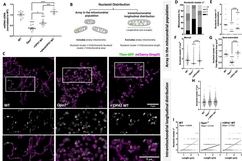
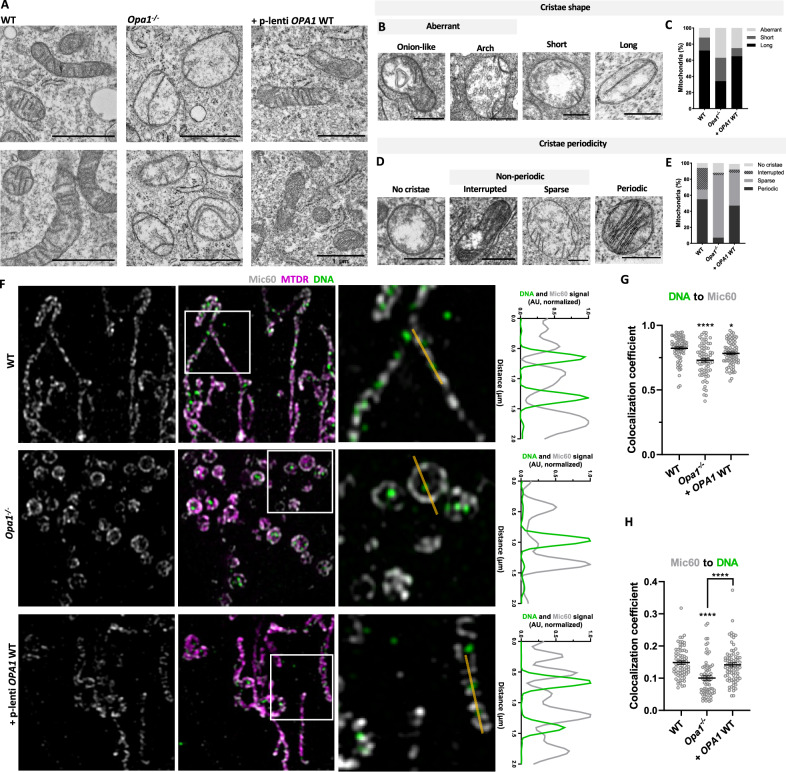
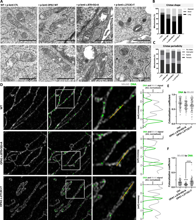
Optic atrophy protein 1 (OPA1) mediates inner mitochondrial membrane (IMM) fusion and cristae organization. Mutations in OPA1 cause autosomal dominant optic atrophy (ADOA), a leading cause of blindness. Cells from ADOA patients show impaired mitochondrial fusion, cristae structure, bioenergetic function, and mitochondrial DNA (mtDNA) integrity. The mtDNA encodes electron transport chain subunits and is packaged into nucleoids spread within the mitochondrial population. Nucleoids interact with the IMM, and their distribution is tightly linked to mitochondrial fusion and cristae shaping. Yet, little is known about the physio-pathological relevance of nucleoid distribution. We studied the effect of OPA1 and ADOA-associated mutants on nucleoid distribution using high-resolution confocal microscopy. We applied a novel model incorporating the mitochondrial context, separating nucleoid distribution into the array in the mitochondrial population and intramitochondrial longitudinal distribution. Opa1-null cells showed decreased mtDNA levels and nucleoid abundance. Also, loss of Opa1 led to an altered distribution of nucleoids in the mitochondrial population, loss of cristae periodicity, and altered nucleoids to cristae proximity partly rescued by OPA1 isoform 1. Overexpression of WT OPA1 or ADOA-causing mutants c.870+5 G > A or c.2713 C > T in WT cells, showed perturbed nucleoid array in the mitochondria population associated with cristae disorganization, which was partly reproduced in Skeletal muscle-derived fibroblasts from ADOA patients harboring the same mutants. Opa1-null and cells overexpressing ADOA mutants accumulated mitochondria without nucleoids. Interestingly, intramitochondrial nucleoid distribution was only altered in Opa1-null cells. Altogether, our results highlight the relevance of OPA1 in nucleoid distribution in the mitochondrial landscape and at a single-organelle level and shed light on new components of ADOA etiology.
视神经萎缩蛋白 1(OPA1)介导线粒体内膜(IMM)融合和嵴结构。OPA1 突变导致常染色体显性视神经萎缩(ADOA),这是失明的主要原因。来自 ADOA 患者的细胞表现出线粒体融合、嵴结构、生物能量功能和线粒体 DNA(mtDNA)完整性受损。mtDNA 编码电子传递链亚基,并被包装成散布在线粒体群中的核体。核体与 IMM 相互作用,其分布与线粒体融合和嵴形成紧密相关。然而,核体分布的生理病理学相关性知之甚少。我们使用高分辨率共聚焦显微镜研究了 OPA1 和 ADOA 相关突变对核体分布的影响。我们应用了一种新的模型,将线粒体环境纳入其中,将核体分布分为线粒体群中的阵列和线粒体内部的纵向分布。Opa1 缺失细胞显示 mtDNA 水平和核体丰度降低。此外,Opa1 的缺失导致核体在线粒体群中的分布改变,嵴周期性丧失,并且核体与嵴的接近度改变,这部分被 OPA1 同工型 1 挽救。在 WT 细胞中过表达 WT OPA1 或 ADOA 致病突变 c.870+5G>A 或 c.2713C>T,显示线粒体群中的核体阵列受到干扰,与嵴结构紊乱有关,这在携带相同突变的 ADOA 患者的骨骼肌衍生成纤维细胞中部分重现。Opa1 缺失和过表达 ADOA 突变体的细胞积累没有核体的线粒体。有趣的是,只有在 Opa1 缺失细胞中才会改变线粒体内部的核体分布。总之,我们的研究结果强调了 OPA1 在线粒体景观和单个细胞器水平的核体分布中的相关性,并揭示了 ADOA 病因的新成分。