• 文献检索
  • 文档翻译
  • 深度研究
  • 学术资讯
  • Suppr Zotero 插件Zotero 插件
  • 邀请有礼
  • 套餐&价格
  • 历史记录
应用&插件
Suppr Zotero 插件Zotero 插件浏览器插件Mac 客户端Windows 客户端微信小程序
定价
高级版会员购买积分包购买API积分包
服务
文献检索文档翻译深度研究API 文档MCP 服务
关于我们
关于 Suppr公司介绍联系我们用户协议隐私条款
关注我们

Suppr 超能文献

核心技术专利:CN118964589B侵权必究
粤ICP备2023148730 号-1Suppr @ 2026

文献检索

告别复杂PubMed语法,用中文像聊天一样搜索,搜遍4000万医学文献。AI智能推荐,让科研检索更轻松。

立即免费搜索

文件翻译

保留排版,准确专业,支持PDF/Word/PPT等文件格式,支持 12+语言互译。

免费翻译文档

深度研究

AI帮你快速写综述,25分钟生成高质量综述,智能提取关键信息,辅助科研写作。

立即免费体验

纳米尺度下的线粒体的光学显微镜观察。

Light Microscopy of Mitochondria at the Nanoscale.

机构信息

Department of NanoBiophotonics, Max Planck Institute for Biophysical Chemistry, 37077 Göttingen, Germany; email:

Clinic of Neurology, University Medical Center Göttingen, 37075 Göttingen, Germany.

出版信息

Annu Rev Biophys. 2020 May 6;49:289-308. doi: 10.1146/annurev-biophys-121219-081550. Epub 2020 Feb 24.

DOI:10.1146/annurev-biophys-121219-081550
PMID:32092283
原文链接:https://pmc.ncbi.nlm.nih.gov/articles/PMC7610798/
Abstract

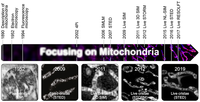
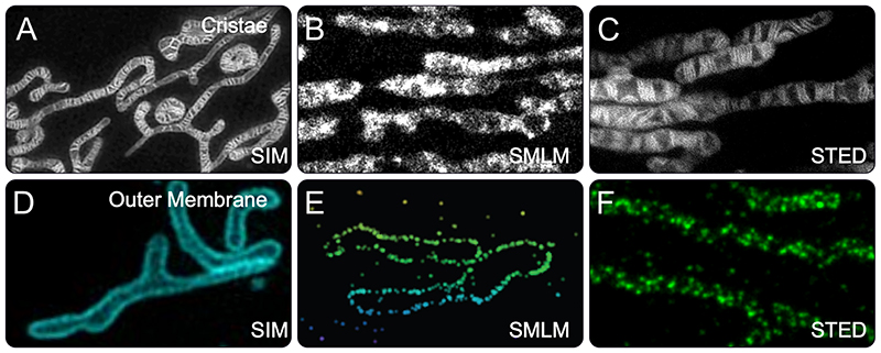
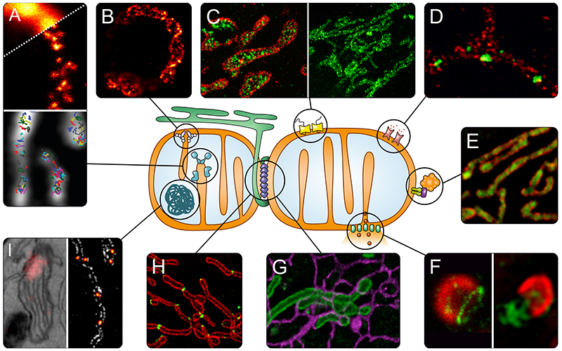
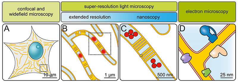
Mitochondria are essential for eukaryotic life. These double-membrane organelles often form highly dynamic tubular networks interacting with many cellular structures. Their highly convoluted contiguous inner membrane compartmentalizes the organelle, which is crucial for mitochondrial function. Since the diameter of the mitochondrial tubules is generally close to the diffraction limit of light microscopy, it is often challenging, if not impossible, to visualize submitochondrial structures or protein distributions using conventional light microscopy. This renders super-resolution microscopy particularly valuable, and attractive, for studying mitochondria. Super-resolution microscopy encompasses a diverse set of approaches that extend resolution, as well as nanoscopy techniques that can even overcome the diffraction limit. In this review, we provide an overview of recent studies using super-resolution microscopy to investigate mitochondria, discuss the strengths and opportunities of the various methods in addressing specific questions in mitochondrial biology, and highlight potential future developments.

摘要

线粒体对于真核生物的生命活动至关重要。这些双层膜细胞器通常形成高度动态的管状网络,与许多细胞结构相互作用。它们高度曲折的连续内膜将细胞器分隔开,这对线粒体的功能至关重要。由于线粒体小管的直径通常接近光显微镜的衍射极限,如果不是不可能的话,使用传统的光显微镜可视化亚线粒体结构或蛋白质分布通常是具有挑战性的。这使得超分辨率显微镜在研究线粒体方面特别有价值和吸引力。超分辨率显微镜包括一系列扩展分辨率的方法,以及可以克服衍射极限的纳米显微镜技术。在这篇综述中,我们提供了使用超分辨率显微镜研究线粒体的最新研究概述,讨论了各种方法在解决线粒体生物学特定问题方面的优势和机会,并强调了潜在的未来发展。

https://cdn.ncbi.nlm.nih.gov/pmc/blobs/53ea/7610798/27b10a3bc325/EMS123370-f004.jpg
https://cdn.ncbi.nlm.nih.gov/pmc/blobs/53ea/7610798/50cb95c3e71d/EMS123370-f001.jpg
https://cdn.ncbi.nlm.nih.gov/pmc/blobs/53ea/7610798/e1c52d376f23/EMS123370-f002.jpg
https://cdn.ncbi.nlm.nih.gov/pmc/blobs/53ea/7610798/1f8a5980a60b/EMS123370-f003.jpg
https://cdn.ncbi.nlm.nih.gov/pmc/blobs/53ea/7610798/27b10a3bc325/EMS123370-f004.jpg

相似文献

1
Light Microscopy of Mitochondria at the Nanoscale.纳米尺度下的线粒体的光学显微镜观察。
Annu Rev Biophys. 2020 May 6;49:289-308. doi: 10.1146/annurev-biophys-121219-081550. Epub 2020 Feb 24.
2
Super-resolution microscopy of mitochondria.线粒体的超分辨率显微镜技术。
Curr Opin Chem Biol. 2014 Jun;20:9-15. doi: 10.1016/j.cbpa.2014.03.019. Epub 2014 Apr 25.
3
Light microscopic analysis of mitochondrial heterogeneity in cell populations and within single cells.细胞群体及单细胞中线粒体异质性的光镜分析。
Adv Biochem Eng Biotechnol. 2011;124:1-19. doi: 10.1007/10_2010_81.
4
STED super-resolution microscopy reveals an array of MINOS clusters along human mitochondria.STED 超分辨率显微镜揭示了人类线粒体中沿 MINOS 簇的排列。
Proc Natl Acad Sci U S A. 2013 May 28;110(22):8936-41. doi: 10.1073/pnas.1301820110. Epub 2013 May 15.
5
Live-cell STED nanoscopy of mitochondrial cristae.活细胞 STED 纳米超分辨显微镜观察线粒体嵴。
Sci Rep. 2019 Aug 27;9(1):12419. doi: 10.1038/s41598-019-48838-2.
6
Application of super-resolution microscopy in mitochondria-dynamic diseases.超分辨率显微镜在线粒体动态疾病中的应用。
Adv Drug Deliv Rev. 2023 Sep;200:115043. doi: 10.1016/j.addr.2023.115043. Epub 2023 Aug 1.
7
Uncovering the impact of UV radiation on mitochondria in dermal cells: a STED nanoscopy study.揭示紫外线辐射对皮肤细胞中线粒体的影响:基于 STED 纳米显微镜的研究。
Sci Rep. 2024 Apr 15;14(1):8675. doi: 10.1038/s41598-024-55778-z.
8
Mitochondrial cristae revealed with focused light.聚焦光显示的线粒体嵴。
Nano Lett. 2009 Jun;9(6):2508-10. doi: 10.1021/nl901398t.
9
Nanoscopy for nanoscience: how super-resolution microscopy extends imaging for nanotechnology.纳米科学中的纳米显微镜:超分辨率显微镜如何为纳米技术扩展成像。
Wiley Interdiscip Rev Nanomed Nanobiotechnol. 2015 May-Jun;7(3):266-81. doi: 10.1002/wnan.1300. Epub 2014 Oct 9.
10
Quantitative dSTORM super-resolution microscopy localizes Aurora kinase A/AURKA in the mitochondrial matrix.定量直接随机光学重建显微镜(dSTORM)超分辨率显微镜将极光激酶A/AURKA定位在线粒体基质中。
Biol Cell. 2021 Nov;113(11):458-473. doi: 10.1111/boc.202100021. Epub 2021 Sep 17.

引用本文的文献

1
Advancements and perspectives on organelle-targeted fluorescent probes for super-resolution SIM imaging.用于超分辨率结构光照明显微成像的细胞器靶向荧光探针的研究进展与展望
Chem Sci. 2025 Sep 8. doi: 10.1039/d5sc04640h.
2
Super-resolution microscopy of mitochondrial mRNAs.线粒体信使核糖核酸的超分辨率显微镜检查
Nat Commun. 2025 Jul 10;16(1):6391. doi: 10.1038/s41467-025-61577-5.
3
A data-driven model for mitochondrial inner membrane remodeling as a driving force of organelle shaping.一种以数据为驱动的线粒体内膜重塑模型,作为细胞器塑形的驱动力。

本文引用的文献

1
Individual cristae within the same mitochondrion display different membrane potentials and are functionally independent.同一线粒体中的个体嵴显示出不同的膜电位,并且在功能上是独立的。
EMBO J. 2019 Nov 15;38(22):e101056. doi: 10.15252/embj.2018101056. Epub 2019 Oct 14.
2
Photoactivation of silicon rhodamines via a light-induced protonation.通过光诱导质子化实现硅罗丹明的光活化。
Nat Commun. 2019 Oct 8;10(1):4580. doi: 10.1038/s41467-019-12480-3.
3
Live-cell STED nanoscopy of mitochondrial cristae.活细胞 STED 纳米超分辨显微镜观察线粒体嵴。
J Cell Sci. 2025 Jun 15;138(12). doi: 10.1242/jcs.263850. Epub 2025 Jun 20.
4
DeepCristae, a CNN for the restoration of mitochondria cristae in live microscopy images.DeepCristae,一种用于在活细胞显微镜图像中恢复线粒体嵴的卷积神经网络。
Commun Biol. 2025 Feb 26;8(1):320. doi: 10.1038/s42003-025-07684-x.
5
The Matrix of Mitochondrial Imaging: Exploring Spatial Dimensions.线粒体成像的基质:探索空间维度
Biomolecules. 2025 Feb 5;15(2):229. doi: 10.3390/biom15020229.
6
Revealing mitochondrial architecture and functions with single molecule localization microscopy.利用单分子定位显微镜揭示线粒体的结构与功能。
Biol Cell. 2025 Jan;117(1):e2400082. doi: 10.1111/boc.202400082.
7
OPA1 and disease-causing mutants perturb mitochondrial nucleoid distribution.OPA1 和致病变异体扰乱了线粒体基因组的分布。
Cell Death Dis. 2024 Nov 30;15(11):870. doi: 10.1038/s41419-024-07165-9.
8
Photobleaching and phototoxicity of mitochondria in live cell fluorescent super-resolution microscopy.活细胞荧光超分辨率显微镜中线粒体的光漂白和光毒性
Mitochondrial Commun. 2024;2:38-47. doi: 10.1016/j.mitoco.2024.03.001. Epub 2024 Mar 16.
9
Visualizing mitochondrial dynamics at the nanoscale.在纳米尺度上可视化线粒体动力学。
Light Sci Appl. 2024 Sep 9;13(1):244. doi: 10.1038/s41377-024-01582-3.
10
Visualization of cristae and mtDNA interactions via STED nanoscopy using a low saturation power probe.通过使用低饱和功率探针的受激发射损耗纳米显微镜观察嵴与线粒体DNA的相互作用。
Light Sci Appl. 2024 May 24;13(1):116. doi: 10.1038/s41377-024-01463-9.
Sci Rep. 2019 Aug 27;9(1):12419. doi: 10.1038/s41598-019-48838-2.
4
A photostable fluorescent marker for the superresolution live imaging of the dynamic structure of the mitochondrial cristae.一种用于超分辨活细胞成像线粒体嵴动态结构的光稳定荧光标记物。
Proc Natl Acad Sci U S A. 2019 Aug 6;116(32):15817-15822. doi: 10.1073/pnas.1905924116. Epub 2019 Jul 23.
5
Mitochondrial fission requires DRP1 but not dynamins.线粒体分裂需要动力相关蛋白1(DRP1),但不需要发动蛋白。
Nature. 2019 Jun;570(7761):E34-E42. doi: 10.1038/s41586-019-1296-y. Epub 2019 Jun 19.
6
Image scanning microscopy.图像扫描显微镜。
Curr Opin Chem Biol. 2019 Aug;51:74-83. doi: 10.1016/j.cbpa.2019.05.011. Epub 2019 Jun 13.
7
Mitochondrial fusion is required for regulation of mitochondrial DNA replication.线粒体融合对于调控线粒体 DNA 复制是必需的。
PLoS Genet. 2019 Jun 6;15(6):e1008085. doi: 10.1371/journal.pgen.1008085. eCollection 2019 Jun.
8
Mic60 exhibits a coordinated clustered distribution along and across yeast and mammalian mitochondria.Mic60 在线粒体中沿酵母和哺乳动物的线粒体呈协调的簇状分布。
Proc Natl Acad Sci U S A. 2019 May 14;116(20):9853-9858. doi: 10.1073/pnas.1820364116. Epub 2019 Apr 26.
9
The Expanding and Unexpected Functions of Mitochondria Contact Sites.线粒体接触位点的不断扩展和意想不到的功能。
Trends Cell Biol. 2019 Jul;29(7):580-590. doi: 10.1016/j.tcb.2019.02.009. Epub 2019 Mar 28.
10
Three-dimensional super-resolution imaging of live whole cells using galvanometer-based structured illumination microscopy.使用基于振镜的结构照明显微镜对活的完整细胞进行三维超分辨率成像。
Opt Express. 2019 Mar 4;27(5):7237-7248. doi: 10.1364/OE.27.007237.