Morin Marie-Christine, D'Astous Jérôme
Centre Vétérinaire Daubigny, Québec City, QC, Canada.
Front Vet Sci. 2024 Nov 15;11:1499465. doi: 10.3389/fvets.2024.1499465. eCollection 2024.
The objectives of the present study were (1) to describe the anatomy of the endodontic system of the dog's maxillary fourth premolar tooth (MxPM4) in relation to the morphology of the crown, (2) to determine if variations of the endodontic system exist, and (3) to look at the implications for endodontic treatment.
Ten MxPM4 were harvested en bloc and scanned using micro-computed tomography (micro-CT).
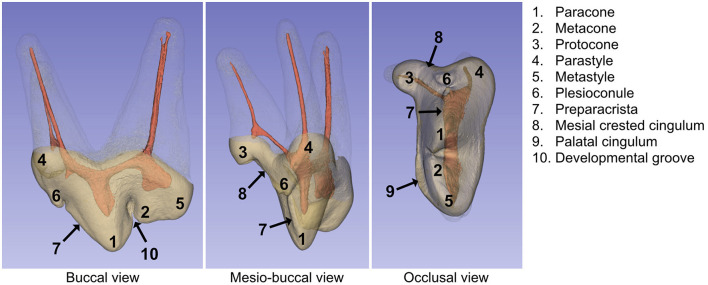
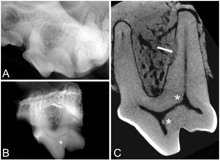
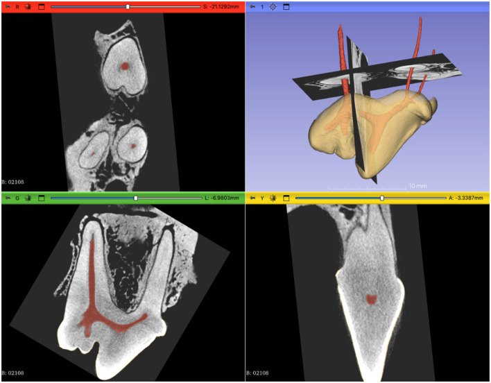
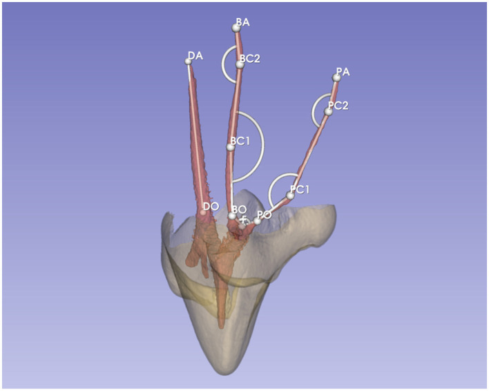
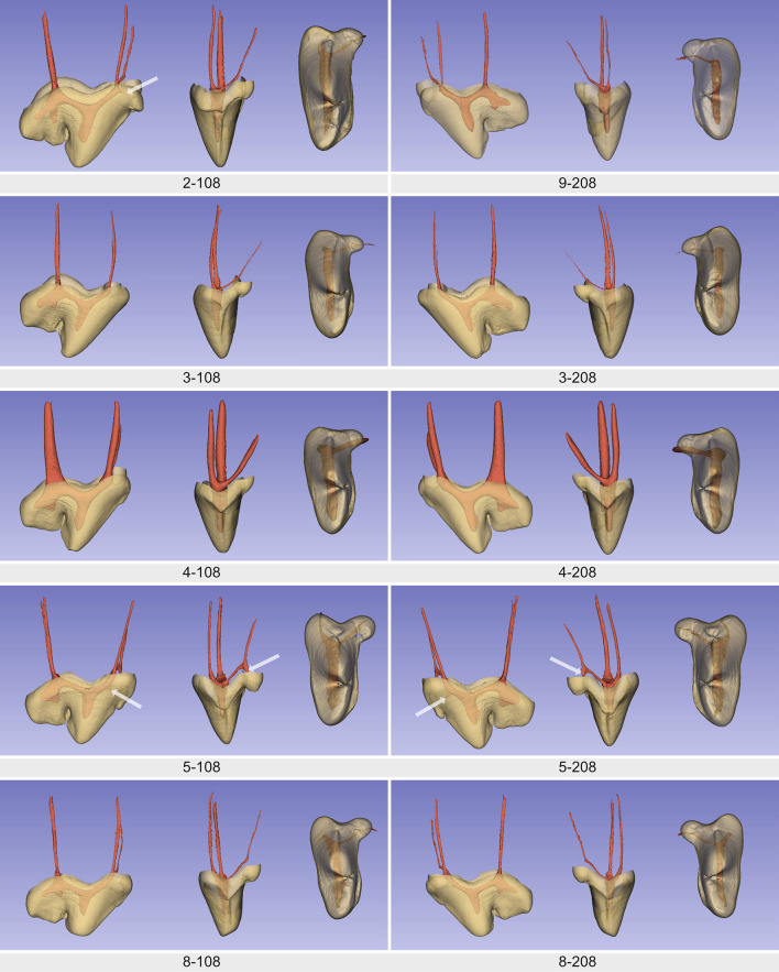
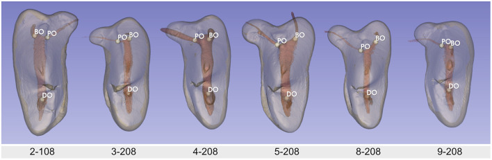
The morphology of the pulp chamber mostly corresponded with the shape of the crown. Three pulp horns were clearly visible and related to the paracone, the metacone, and the metastyle. Nevertheless, the pulp horns of the metacone and metastyle could be fused, partially fused or distinct. Other pulp projections were also present, but rarely, in the parastyle, the protocone, and the plesioconule. All teeth showed a noticeable angulation of an average of 150 degrees at the coronal third of the mesiopalatal canal.
Thus, the most common transcoronal approach for root canal treatment does not allow a straight access to the apex. There were also minor variations in the locations of the canal orifices. This first micro-CT study of the MxPM4 in dogs showed anatomical features and variations of the pulp cavity that have not been described before.
本研究的目的是:(1)描述犬上颌第四前磨牙(MxPM4)牙髓系统的解剖结构及其与牙冠形态的关系;(2)确定牙髓系统是否存在变异;(3)探讨这些变异对牙髓治疗的影响。
完整采集10颗MxPM4牙齿,采用显微计算机断层扫描(micro-CT)进行扫描。
髓腔形态大多与牙冠形状相符。可见三个髓角,分别与副尖、主尖和后尖相对应。然而,主尖和后尖的髓角可能融合、部分融合或分离。在副尖、原尖和近中副小尖处也存在其他髓突,但较少见。所有牙齿在近中腭根管冠方三分之一处均有明显的平均150度的角度。
因此,最常用的根管治疗经冠部入路无法直接到达根尖。根管口的位置也存在微小变异。这项对犬MxPM4的首次显微CT研究显示了以前未描述过的牙髓腔解剖特征和变异。