Dodina Maria, Gurtsieva Dzerassa, Karabelsky Alexander, Minskaia Ekaterina
Sirius University of Science and Technology, Krasnodar, Russia.
Front Cell Dev Biol. 2024 Nov 15;12:1455140. doi: 10.3389/fcell.2024.1455140. eCollection 2024.
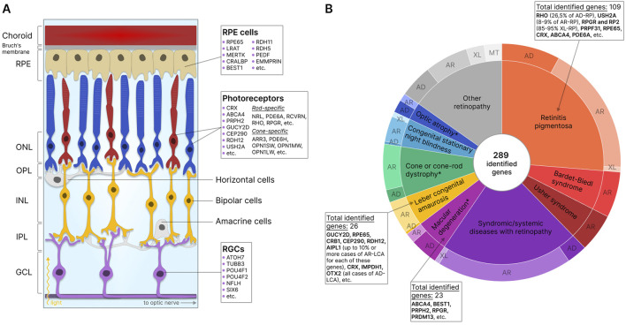
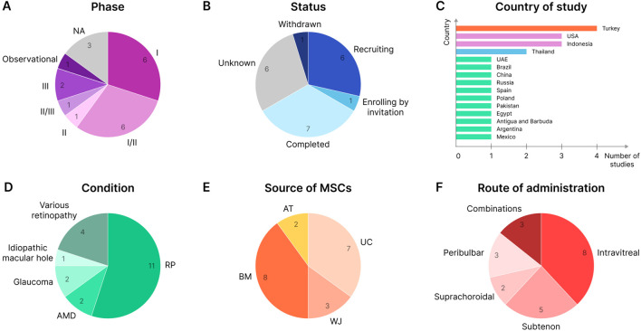
Retinal pathologies are major causes of vision impairment and blindness in humans, and inherited retinal diseases (IRDs), such as retinitis pigmentosa, Leber congenital amaurosis, and Stargardt disease, greatly contribute to this problem. disease modeling can be used for understanding the development of pathology and for screening therapeutic pharmaceutical compounds. In the preclinical research phase, models complement models by reducing animal studies, decreasing costs, and shortening research timelines. Additionally, animal models may not always accurately replicate the human disease phenotype. This review examines the types of cells that can be used to create IRD models, including retina-specific cell lines, primary retinal cells, induced pluripotent stem cells (iPSCs), and more. Special attention is given to mesenchymal stem cells (MSCs), which are characterized by various isolation sources, relative ease of isolation, and straightforward differentiation. MSCs derived from bone marrow (BM), adipose tissue (AT), dental tissue (DT), umbilical cord (UC), and other sources can differentiate into retinal cells, including photoreceptor cells and retinal pigment epithelial (RPE) cells, dysfunction of which is most commonly associated with IRDs. Subsequent differentiation of MSCs into retinal cells can be carried out via various methods: culturing in induction media supplemented with certain growth factors, co-culturing with retinal cells or in their conditioned media, or regulating gene expression with viral vector-delivered transcription factors (TFs) or microRNAs (miRNAs). Compared to the popular iPSCs, for example, MSC-based models are significantly cheaper and faster to obtain, making them more feasible for large-scale drug screening. Nevertheless, the existing differentiation methods need further optimization for this promising platform to receive the success it deserves.
视网膜病变是人类视力障碍和失明的主要原因,而遗传性视网膜疾病(IRD),如色素性视网膜炎、莱伯先天性黑蒙和斯塔加特病,在很大程度上导致了这一问题。疾病建模可用于了解病理发展过程并筛选治疗性药物化合物。在临床前研究阶段,模型通过减少动物研究、降低成本和缩短研究时间来补充模型。此外,动物模型可能并不总是能准确复制人类疾病表型。本综述探讨了可用于创建IRD模型的细胞类型,包括视网膜特异性细胞系、原代视网膜细胞、诱导多能干细胞(iPSC)等。特别关注间充质干细胞(MSC),其特点是分离来源多样、相对易于分离且分化过程简单。源自骨髓(BM)、脂肪组织(AT)、牙齿组织(DT)、脐带(UC)和其他来源的MSC可分化为视网膜细胞,包括光感受器细胞和视网膜色素上皮(RPE)细胞,其功能障碍最常与IRD相关。随后,MSC向视网膜细胞的分化可通过多种方法进行:在添加特定生长因子的诱导培养基中培养、与视网膜细胞共培养或在其条件培养基中培养,或用病毒载体递送的转录因子(TF)或微小RNA(miRNA)调节基因表达。例如,与流行的iPSC相比,基于MSC的模型获取成本显著更低且速度更快,使其在大规模药物筛选中更具可行性。然而,对于这个有前景的平台而言,现有的分化方法需要进一步优化,以获得应有的成功。