Bischof Gérard N, Jaeger Elena, Giehl Kathrin, Jessen Frank, Onur Oezguer A, O'Bryant Sid, Kara Esra, Weiss Peter H, Drzezga Alexander
From the Multimodal Neuroimaging Group, Department of Nuclear Medicine (G.N.B., E.J., K.G., A.D.), Department of Psychiatry (F.J.), Department of Neurology (O.A.O., E.K., P.H.W.), Medical Faculty and University Hospital of Cologne, University of Cologne; Molecular Organization of the Brain (G.N.B., A.D.), Institute for Neuroscience and Medicine II, Research Center Juelich; German Center for Neurodegenerative Diseases (F.J.), Bonn/Cologne, Germany; Institute for Translational Research (S.O.B.), and Department of Family Medicine (S.O.B.), Texas College of Osteopathic Medicine, University of North Texas Health Science Center, Fort Worth; and Cognitive Neuroscience (P.H.W.), Institute for Neuroscience and Medicine (INM-3), Research Center Juelich, Germany.
Neurology. 2024 Dec 24;103(12):e210062. doi: 10.1212/WNL.0000000000210062. Epub 2024 Dec 3.
Apraxia is a frequently observed symptom in Alzheimer disease (AD), but the causal pathomechanism underlying this dysfunction is not well understood. Previous studies have demonstrated associations between various cognitive dysfunctions in AD and cortical tau deposition in specific brain areas, suggesting a causal relationship. Thus, we hypothesized that specific regional patterns of tau pathology in praxis-related brain regions may be associated with apraxic deficits in AD. For this purpose, we performed PET imaging with the second-generation tau-PET tracer [18F]PI-2620 in a well-defined group of patients with AD (N = 33) who had been systematically assessed for apraxia.
Patients with a biomarker-confirmed diagnosis of AD were recruited in addition to a sample of cognitively unimpaired (CU) control participants. Both groups underwent apraxia assessments with the Dementia Apraxia Screening Test. In addition, PET imaging with [18F]PI-2620 was performed to assess tau pathology in the patients with AD. To specifically investigate the association of apraxia severity with regional tau pathology, we compared the PET data from this group with an independent sample of amyloid-negative cognitively intact participants (CU) by generation of -score deviation maps and submitted these maps to a voxel-based multiple regression analysis.
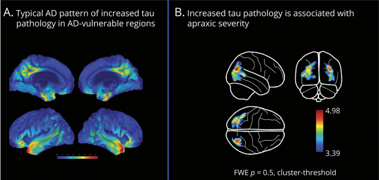
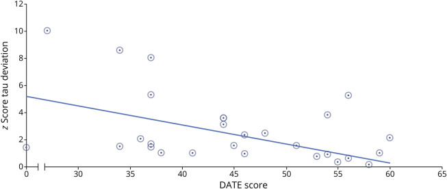
A total of 120 participants (39% female) with a mean age of 67.9 (9.2) years were included in the study (AD = 33; CU; N = 33; CU; N = 54). We identified a significant correlation between circumscribed clusters of tau aggregation in praxis-related brain regions (including parietal (angular gyrus), temporal, and occipital regions) and severity of apraxia in patients with AD. By contrast, no significant correlations between tau tracer uptake in primary motor cortex or subcortical brain regions and apraxia were observed.
These results suggest that tau deposition in specific cortical praxis-related brain regions may induce local neuronal dysfunction leading to a dose-dependent functional decline in praxis performance in AD. The awareness of this relationship could further refine a differentiated individual diagnostic characterization and classification of patients with AD.
失用症是阿尔茨海默病(AD)中常见的症状,但这种功能障碍背后的致病病理机制尚不清楚。先前的研究表明,AD中的各种认知功能障碍与特定脑区的皮质tau沉积之间存在关联,提示存在因果关系。因此,我们假设与运用相关脑区的特定区域tau病理模式可能与AD中的失用症缺陷有关。为此,我们使用第二代tau-PET示踪剂[18F]PI-2620对一组经过系统失用症评估的明确AD患者(N = 33)进行了PET成像。
除了认知未受损(CU)的对照参与者样本外,还招募了生物标志物确诊的AD患者。两组均使用痴呆失用症筛查测试进行失用症评估。此外,对AD患者进行了[18F]PI-2620的PET成像以评估tau病理。为了具体研究失用症严重程度与区域tau病理的关联,我们通过生成z分数偏差图将该组的PET数据与淀粉样蛋白阴性认知完整参与者(CU)的独立样本进行比较,并将这些图提交给基于体素的多元回归分析。
共有120名参与者(39%为女性)纳入研究,平均年龄为67.9(9.2)岁(AD = 33;CU;N = 33;CU;N = 54)。我们发现与运用相关脑区(包括顶叶(角回)、颞叶和枕叶区域)中tau聚集的局限性簇与AD患者的失用症严重程度之间存在显著相关性。相比之下,在初级运动皮层或皮质下脑区的tau示踪剂摄取与失用症之间未观察到显著相关性。
这些结果表明,特定皮质运用相关脑区的tau沉积可能诱导局部神经元功能障碍,导致AD中运用能力呈剂量依赖性功能下降。认识到这种关系可以进一步完善AD患者的差异化个体诊断特征和分类。