Papasavvas Ioannis, Tucker William R, Mantovani Alessandro, Fabozzi Lorenzo, Herbort Carl P
Centre for Ophthalmic Specialised Care (COS), Lausanne, Switzerland.
Moorfields Eye Hospital NHS Trust, London, UK.
J Ophthalmic Inflamm Infect. 2024 Dec 4;14(1):63. doi: 10.1186/s12348-024-00442-w.
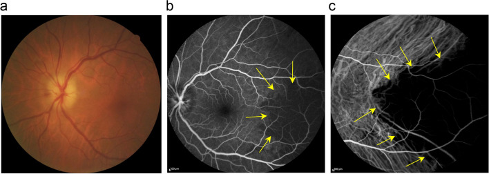
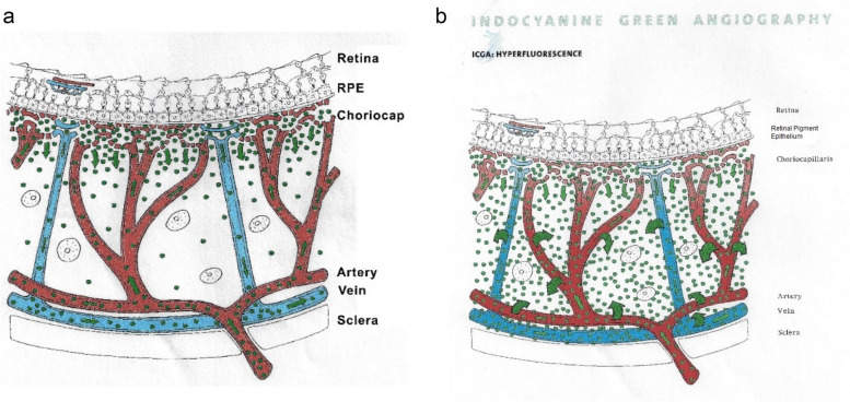
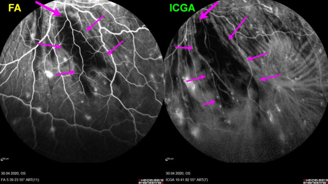
Indocyanine green angiography (ICGA) is the gold standard to diagnose, evaluate and follow up choroidal inflammation. It allows clinicians to precisely determine the type and extension of choroidal vasculitis in the two main choroidal structures, the choriocapillaris and the choroidal stroma. The presence of choroidal vasculitis is often overlooked by the physician who often does not include ICGA in the investigation of posterior uveitis.
To describe choroidal vasculitis by analysing its ICGA signs in order to investigate and follow choroiditis and determine the pathophysiological mechanisms of inflammation of choroidal vessels.
The tutorial is presenting the normal findings in a non-inflamed choroid and the semiology of diverse choroidal vasculitis conditions, followed by practical illustrations using typical cases.
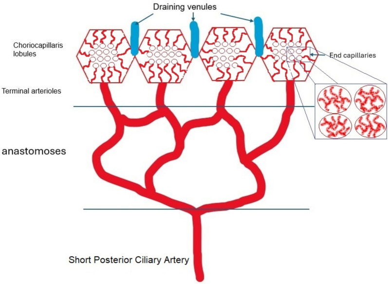
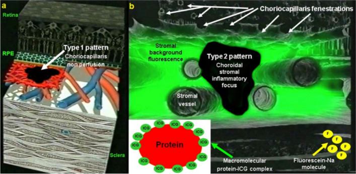
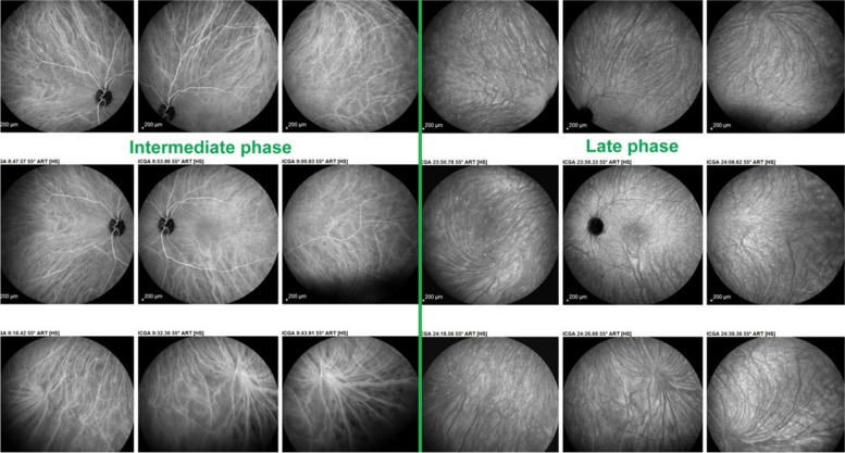
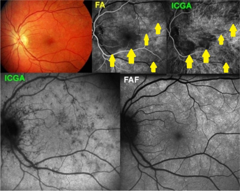
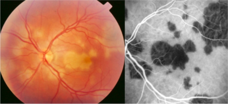
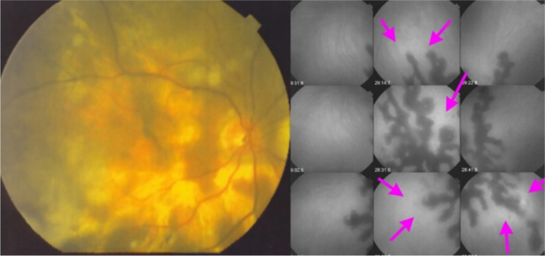
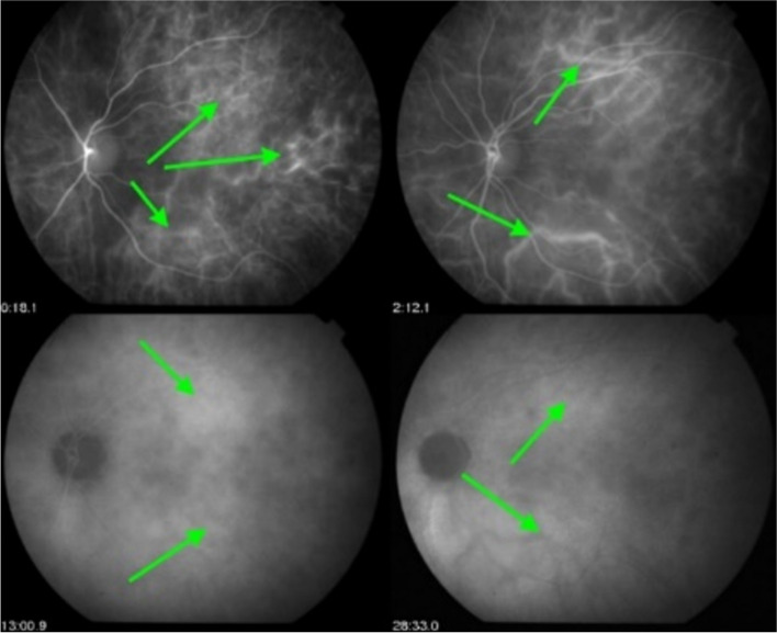
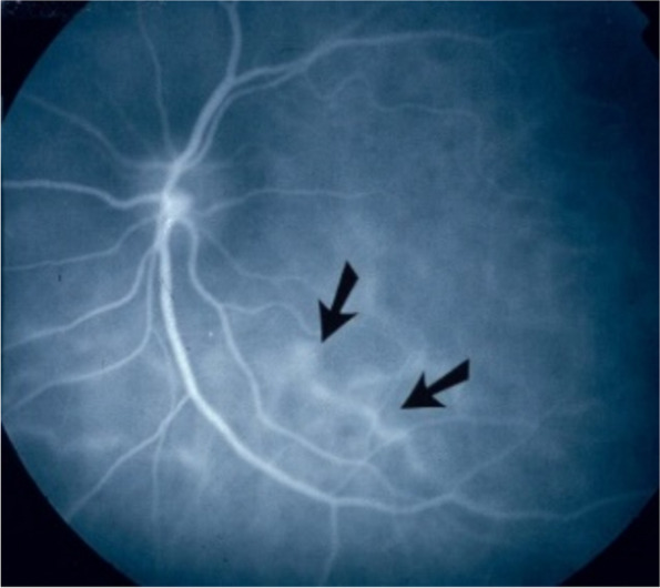
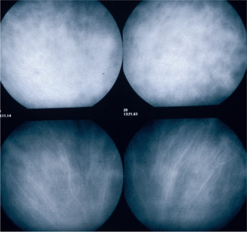
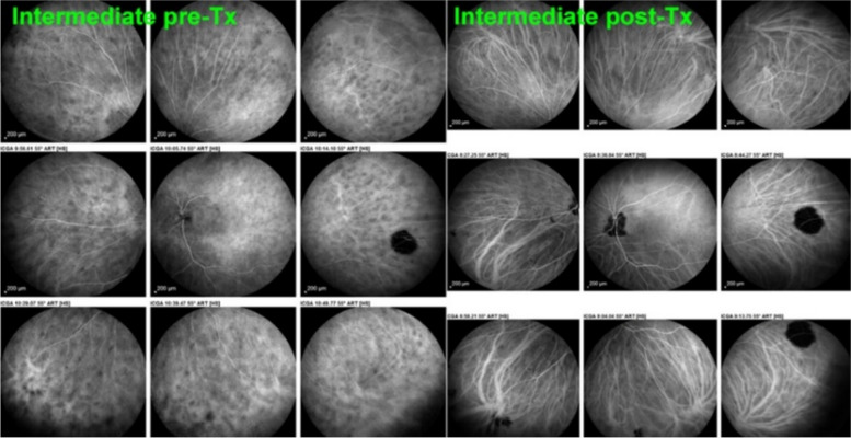
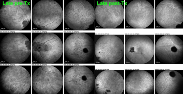
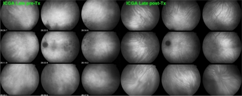
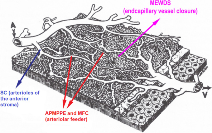
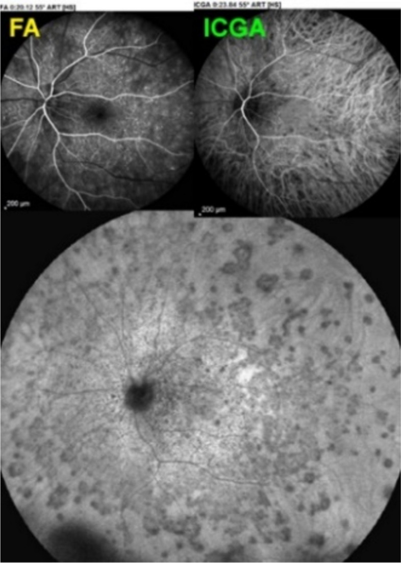
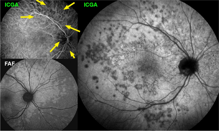
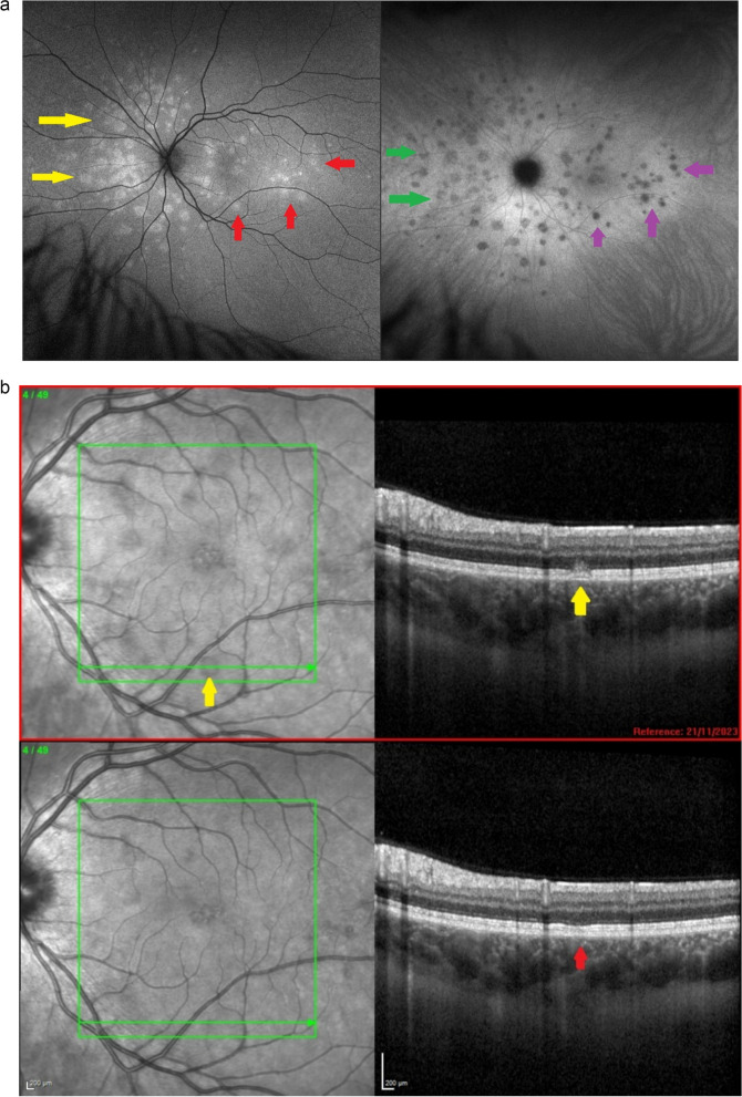
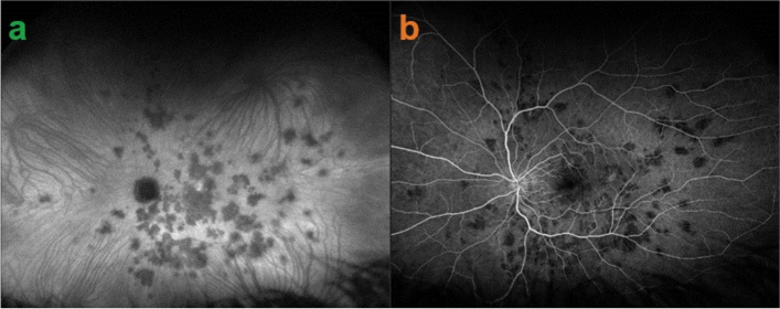
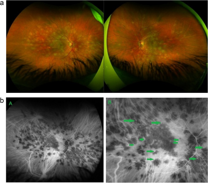
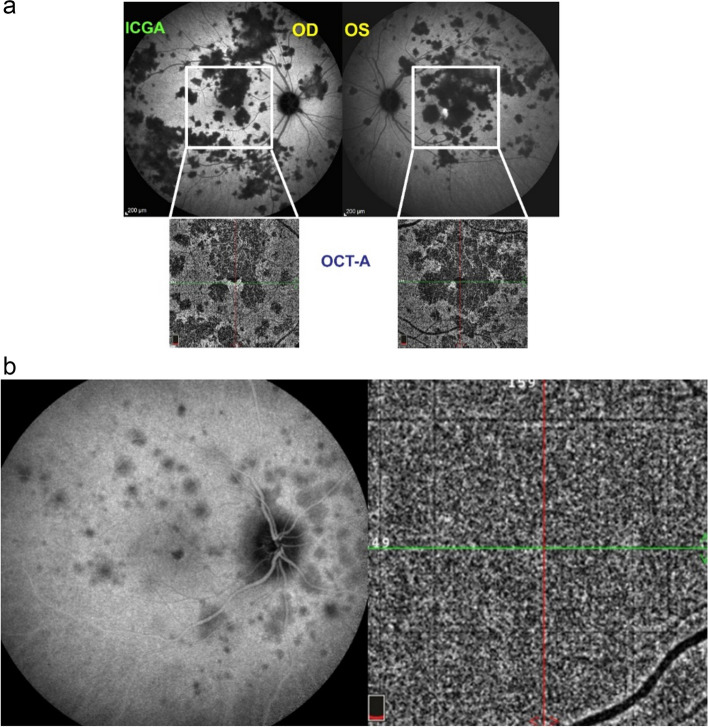
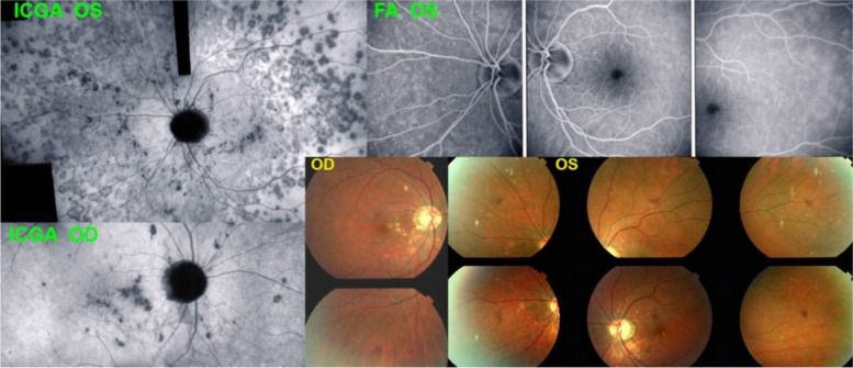
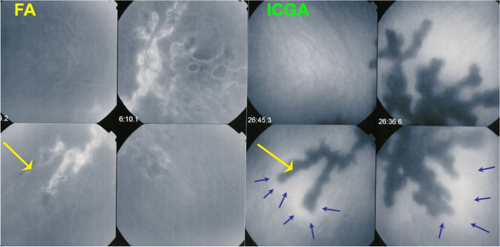
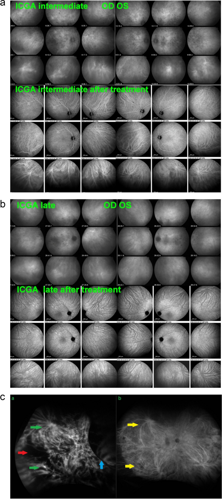
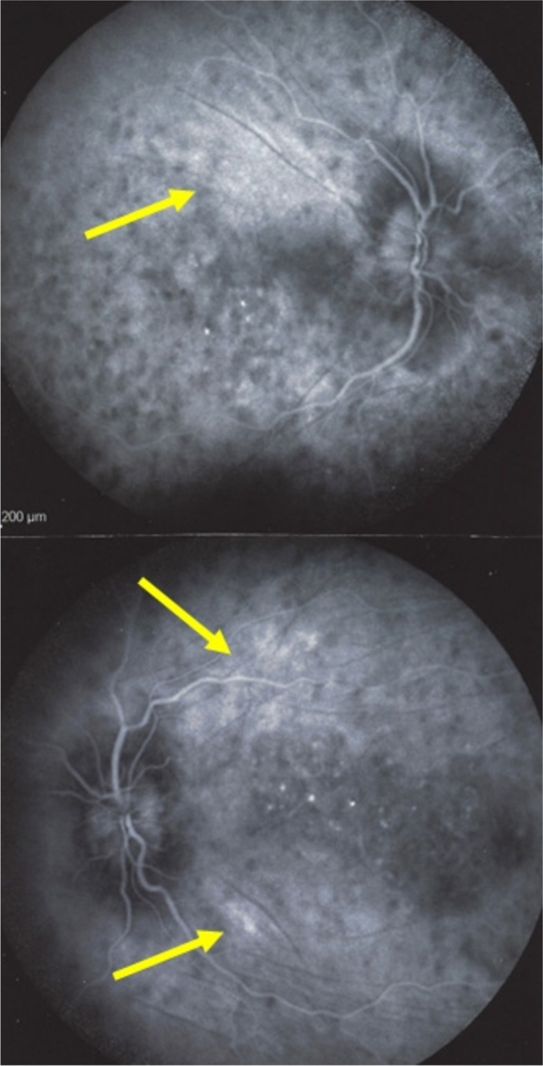
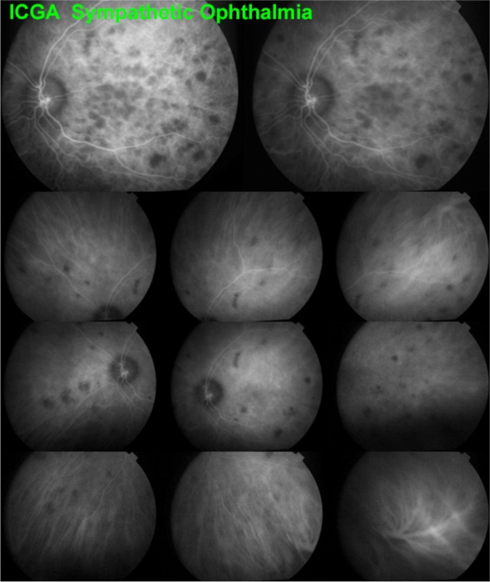
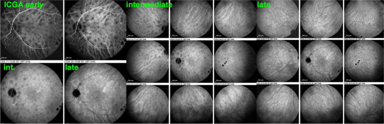
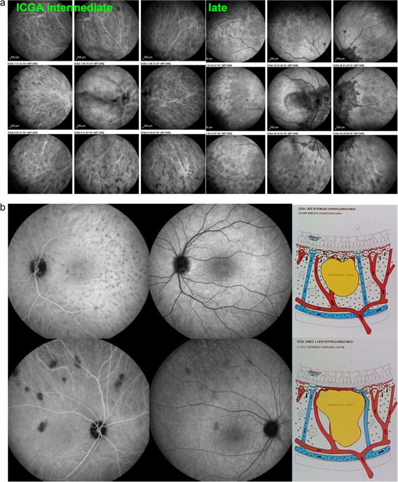
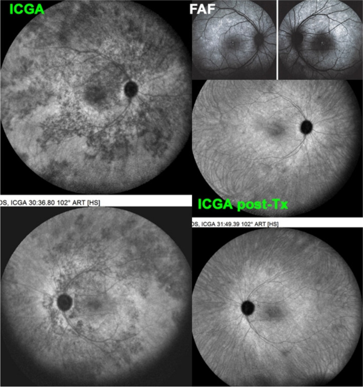
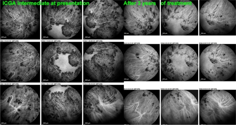
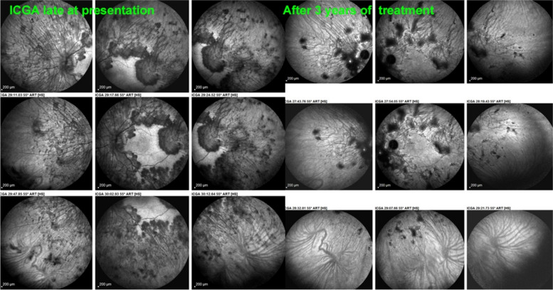
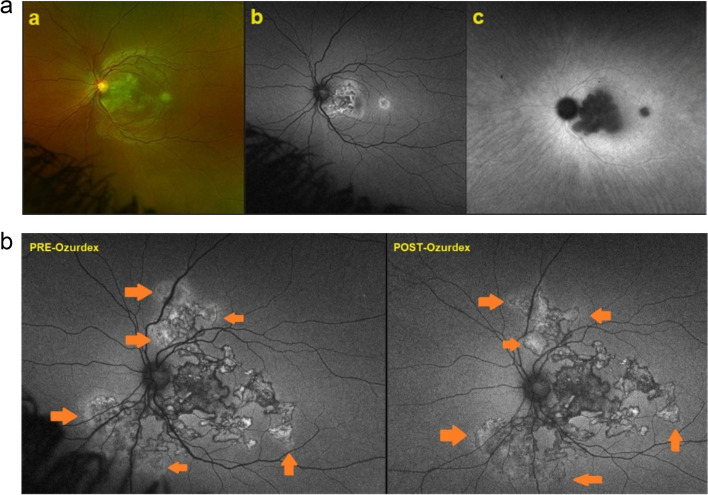
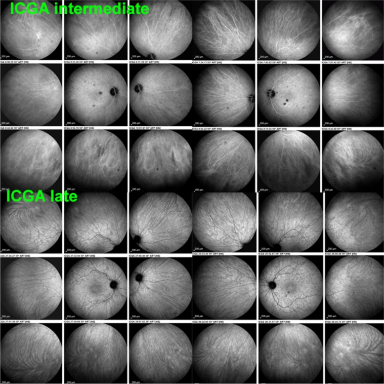
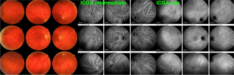
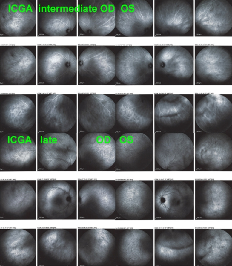
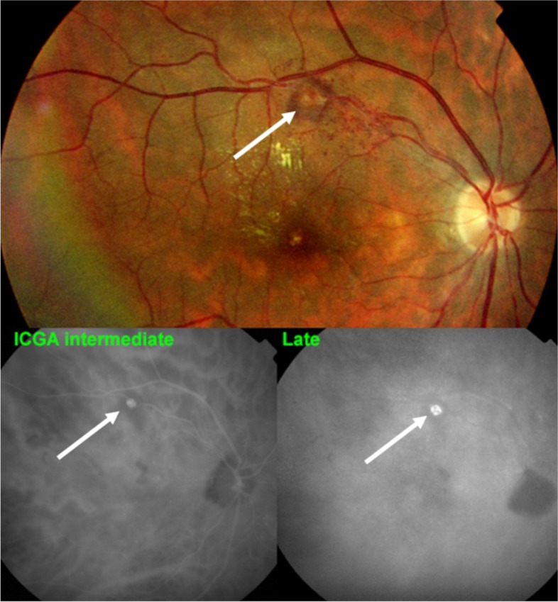
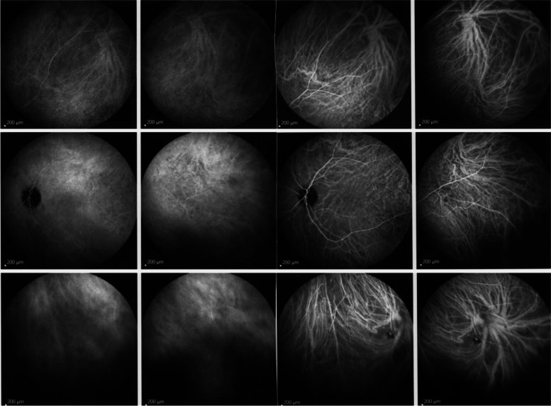
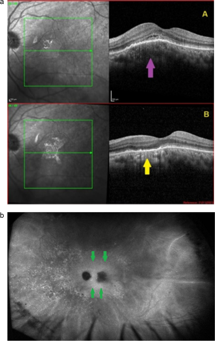
The two identified patterns of choroidal vasculitis corresponded on one side to choriocapillaritis appearing as areas of hypofluorescence depicting the involvement and extension of choriocapillaris inflammatory non-perfusion. The vasculitis of the choriocapillaris goes from limited and reversible when distal endcapillary vessels are involved such as in Multiple Evanescent White Dot Syndrome (MEWDS) to more severe involvement in Acute Posterior Multifocal Placoid Pigment Epitheliopathy (APMPPE), Multifocal Choroiditis (MFC) or Serpiginous Choroiditis (SC) with more pronounced non-perfusion causing scars if not treated diligently. On the other side, stromal choroidal vasculitis is characterised by leaking hyperfluorescent vessels that appear fuzzy and at the origin of late diffuse choroidal hyperfluorescence.
Choroidal vasculitis is present in almost all patients with inflammatory choroidal involvement, occlusive in case of choriocapillaritis and leaky in stromal choroiditis causing vessel hyperfluorescence, fuzziness of the choroidal vessels and late diffuse stromal hyperfluorescence on ICGA. Systemic vasculitis entities produce occlusive vasculitis of large choroidal vessels.
吲哚菁绿血管造影(ICGA)是诊断、评估和随访脉络膜炎症的金标准。它使临床医生能够精确确定脉络膜血管炎在脉络膜两个主要结构,即脉络膜毛细血管层和脉络膜基质中的类型和范围。脉络膜血管炎的存在常常被医生忽视,他们在后部葡萄膜炎的检查中往往不进行ICGA检查。
通过分析ICGA征象来描述脉络膜血管炎,以研究和随访脉络膜炎,并确定脉络膜血管炎症的病理生理机制。
本教程展示了非炎症性脉络膜的正常表现以及各种脉络膜血管炎情况的症状学,随后使用典型病例进行实际说明。
确定的两种脉络膜血管炎模式,一方面对应于脉络膜毛细血管炎,表现为低荧光区域,描绘了脉络膜毛细血管层炎症性无灌注的累及范围和程度。脉络膜毛细血管层血管炎从累及远端毛细血管时的局限性和可逆性,如在多发性一过性白点综合征(MEWDS)中,到急性后极部多灶性鳞状色素上皮病变(APMPPE)、多灶性脉络膜炎(MFC)或匐行性脉络膜炎(SC)中更严重的累及,若不及时治疗,更明显的无灌注会导致瘢痕形成。另一方面,基质性脉络膜血管炎的特征是渗漏的高荧光血管,看起来模糊不清,是晚期弥漫性脉络膜高荧光的起源。
几乎所有有炎症性脉络膜受累的患者都存在脉络膜血管炎,脉络膜毛细血管炎时为闭塞性,基质性脉络膜炎时为渗漏性,导致ICGA上血管高荧光、脉络膜血管模糊不清和晚期弥漫性基质高荧光。系统性血管炎实体可导致大脉络膜血管的闭塞性血管炎。