• 文献检索
  • 文档翻译
  • 深度研究
  • 学术资讯
  • Suppr Zotero 插件Zotero 插件
  • 邀请有礼
  • 套餐&价格
  • 历史记录
应用&插件
Suppr Zotero 插件Zotero 插件浏览器插件Mac 客户端Windows 客户端微信小程序
定价
高级版会员购买积分包购买API积分包
服务
文献检索文档翻译深度研究API 文档MCP 服务
关于我们
关于 Suppr公司介绍联系我们用户协议隐私条款
关注我们

Suppr 超能文献

核心技术专利:CN118964589B侵权必究
粤ICP备2023148730 号-1Suppr @ 2026

文献检索

告别复杂PubMed语法,用中文像聊天一样搜索,搜遍4000万医学文献。AI智能推荐,让科研检索更轻松。

立即免费搜索

文件翻译

保留排版,准确专业,支持PDF/Word/PPT等文件格式,支持 12+语言互译。

免费翻译文档

深度研究

AI帮你快速写综述,25分钟生成高质量综述,智能提取关键信息,辅助科研写作。

立即免费体验

多排螺旋CT、牙科锥形束CT和术中三维C型臂在面颅骨区域有效辐射剂量的比较

Comparison of the Effective Radiation Dose in the Region of the Facial Skull Between Multidetector CT, Dental Conebeam CT and Intraoperative 3D C-Arms.

作者信息

Pietzka Sebastian, Grieser Anne, Winter Karsten, Schramm Alexander, Metzger Marc, Semper-Hogg Wiebke, Grunert Michael, Ebeling Marcel, Sakkas Andreas, Wilde Frank

机构信息

Department of Cranio-Maxillo-Facial-Surgery, University Hospital Ulm, Ulm, Germany.

Department of Cranio-Maxillo-Facial-Surgery, German Armed Forces Hospital Ulm, Ulm, Germany.

出版信息

Craniomaxillofac Trauma Reconstr. 2024 Dec;17(4):270-278. doi: 10.1177/19433875231213906. Epub 2023 Nov 10.

DOI:10.1177/19433875231213906
PMID:39634571
原文链接:https://pmc.ncbi.nlm.nih.gov/articles/PMC11613283/
Abstract

STUDY DESIGN

Experimental single-centre study of X-ray absorption using a phantom skull.

OBJECTIVE

This experimental study aimed to compare the radiation doses of different 3D imaging devices used in maxillofacial surgery, including one Multidetector CT (MDCT), two Conebeam CT (CBCT) and four intraoperative 3D C-arms.

METHODS

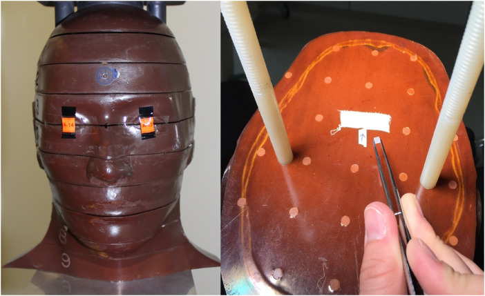
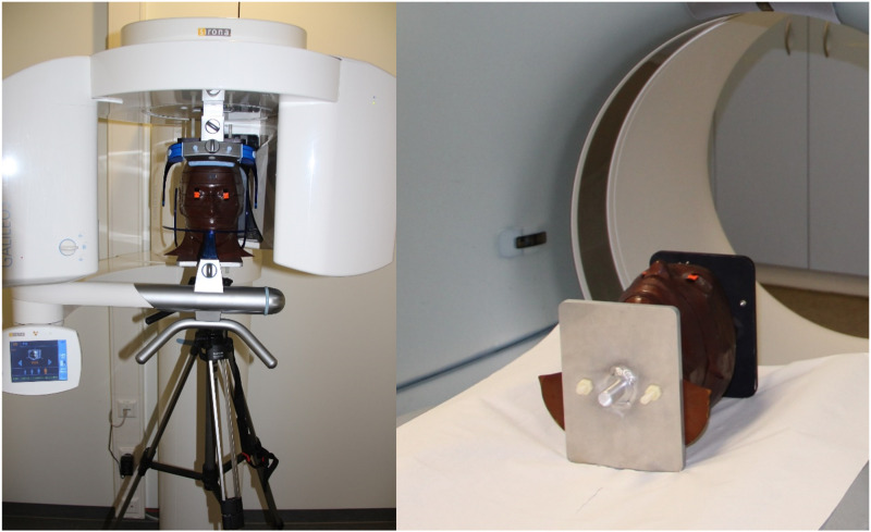
Thermoluminescent dosimeters (TLD) were used to determine the absorbed radiation in an Alderson-Rando phantom skull. The phantom skull was positioned in the before mentioned seven devices and a defined 3D facial skull image was acquired. Subsequently, the TLD'S were read out and the effective doses (ED) and the organ doses (OD) were calculated and compared.

RESULTS

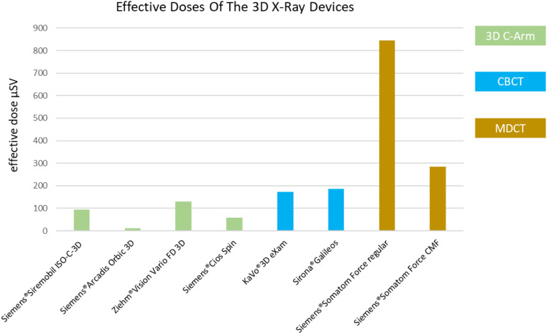
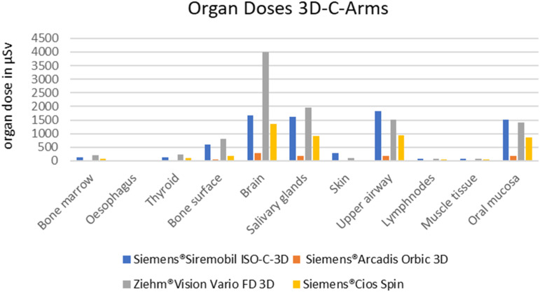
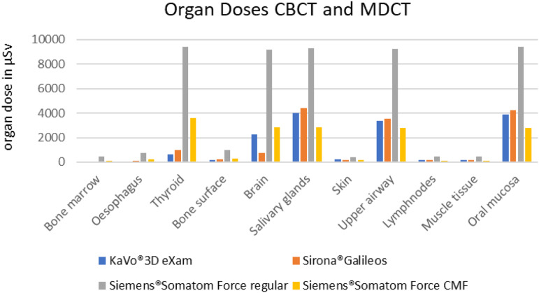
OD varied significantly between tissues as well as between the 3D X-ray devices. The OD of the 3D C-arms were significantly lower than those of all other devices. The OD of the CT, especially in the standard setting, was the highest. Only by special adjustments of the scan protocol regarding CMF requirements for traumatology, the MDCT could achieve almost equivalent doses as the two tested CBCT-scanners. The calculated effective doses were also lowest for the 3D C-arm devices (11.2 to 129.9 μSv). The ED of the MDCT were significant higher (284.52-844.97 μSv) than in all other devices. The ED of the CBCTs (173.7-184.9) were lower than for MDCT but still higher than those of the 3D C-arms.

CONCLUSIONS

Intraoperative imaging using 3D C-arm devices is an effective method to verify reduction results in maxillofacial surgery intraoperatively with significantly lower ED than postoperatively CBCT and MDCT imaging.

摘要

研究设计

使用仿真颅骨进行X射线吸收的单中心实验研究。

目的

本实验研究旨在比较颌面外科中使用的不同3D成像设备的辐射剂量,包括一台多排螺旋CT(MDCT)、两台锥形束CT(CBCT)和四台术中3D C形臂。

方法

使用热释光剂量计(TLD)测定Alderson-Rando仿真颅骨中的吸收辐射。将仿真颅骨放置在上述七种设备中,并获取定义的3D面部颅骨图像。随后,读出TLD并计算和比较有效剂量(ED)和器官剂量(OD)。

结果

OD在组织之间以及3D X射线设备之间存在显著差异。3D C形臂的OD显著低于所有其他设备。CT的OD最高,尤其是在标准设置下。只有通过根据创伤学的CMF要求对扫描协议进行特殊调整,MDCT才能达到与两台测试的CBCT扫描仪几乎相同的剂量。计算得出的有效剂量在3D C形臂设备中也是最低的(11.2至129.9μSv)。MDCT的ED显著高于所有其他设备(284.52 - 844.97μSv)。CBCT的ED(173.7 - 184.9)低于MDCT,但仍高于3D C形臂。

结论

在颌面外科手术中,使用3D C形臂设备进行术中成像,是一种有效验证复位结果的方法,其有效剂量明显低于术后CBCT和MDCT成像。

https://cdn.ncbi.nlm.nih.gov/pmc/blobs/1d55/11613283/289beed5d717/10.1177_19433875231213906-fig5.jpg
https://cdn.ncbi.nlm.nih.gov/pmc/blobs/1d55/11613283/132d9eb73a1f/10.1177_19433875231213906-fig1.jpg
https://cdn.ncbi.nlm.nih.gov/pmc/blobs/1d55/11613283/f3a847b2213e/10.1177_19433875231213906-fig2.jpg
https://cdn.ncbi.nlm.nih.gov/pmc/blobs/1d55/11613283/710a25d1cf3b/10.1177_19433875231213906-fig3.jpg
https://cdn.ncbi.nlm.nih.gov/pmc/blobs/1d55/11613283/8bdd2852e4d8/10.1177_19433875231213906-fig4.jpg
https://cdn.ncbi.nlm.nih.gov/pmc/blobs/1d55/11613283/289beed5d717/10.1177_19433875231213906-fig5.jpg

相似文献

1
Comparison of the Effective Radiation Dose in the Region of the Facial Skull Between Multidetector CT, Dental Conebeam CT and Intraoperative 3D C-Arms.多排螺旋CT、牙科锥形束CT和术中三维C型臂在面颅骨区域有效辐射剂量的比较
Craniomaxillofac Trauma Reconstr. 2024 Dec;17(4):270-278. doi: 10.1177/19433875231213906. Epub 2023 Nov 10.
2
Two examples of indication specific radiation dose calculations in dental CBCT and Multidetector CT scanners.牙科锥形束计算机断层扫描(CBCT)和多层螺旋计算机断层扫描(MDCT)扫描仪中特定适应症辐射剂量计算的两个示例。
Phys Med. 2017 Sep;41:71-77. doi: 10.1016/j.ejmp.2017.03.027. Epub 2017 Apr 6.
3
Evaluation of different low-dose multidetector CT and cone beam CT protocols in maxillary sinus imaging: part I-an in vitro study.不同低剂量多层螺旋CT和锥形束CT方案在上颌窦成像中的评估:第一部分——一项体外研究
Dentomaxillofac Radiol. 2017 Aug;46(6):20160323. doi: 10.1259/dmfr.20160323. Epub 2017 Apr 12.
4
Comparison of effective dose for imaging of mandible between multi-detector CT and cone-beam CT.多排螺旋CT与锥形束CT在下颌骨成像有效剂量的比较。
Imaging Sci Dent. 2012 Jun;42(2):65-70. doi: 10.5624/isd.2012.42.2.65. Epub 2012 Jun 25.
5
Radiation dose evaluation in tomosynthesis and C-arm cone-beam CT examinations with an anthropomorphic phantom.在体层合成摄影术和 C 臂锥形束 CT 检查中用人形体模进行辐射剂量评估。
Med Phys. 2010 Aug;37(8):4298-306. doi: 10.1118/1.3465045.
6
Effective radiation dose of a MSCT, two CBCT and one conventional radiography device in the ankle region.多层螺旋CT、两台锥形束CT和一台传统X线摄影设备在踝关节区域的有效辐射剂量。
J Foot Ankle Res. 2015 Mar 12;8:8. doi: 10.1186/s13047-015-0067-8. eCollection 2015.
7
Cone beam CT paranasal sinuses versus standard multidetector and low dose multidetector CT studies.锥形束CT鼻窦成像与标准多层螺旋CT及低剂量多层螺旋CT研究对比
Am J Otolaryngol. 2016 Jan-Feb;37(1):59-64. doi: 10.1016/j.amjoto.2015.08.002. Epub 2015 Aug 24.
8
Absorbed and effective doses from cone beam volumetric imaging for implant planning.用于种植体规划的锥束容积成像的吸收剂量和有效剂量。
Dentomaxillofac Radiol. 2009 Feb;38(2):79-85. doi: 10.1259/dmfr/14769929.
9
Comparative dosimetry of dental CBCT devices and 64-slice CT for oral and maxillofacial radiology.牙科锥形束计算机断层扫描(CBCT)设备与64层CT在口腔颌面放射学中的剂量学比较
Oral Surg Oral Med Oral Pathol Oral Radiol Endod. 2008 Jul;106(1):106-14. doi: 10.1016/j.tripleo.2008.03.018. Epub 2008 May 27.
10
Cone beam computed tomography for dental and maxillofacial imaging: technique improvement and low-dose protocols.锥形束计算机断层扫描在口腔颌面医学影像学中的应用:技术改进与低剂量方案
Radiol Med. 2017 Aug;122(8):581-588. doi: 10.1007/s11547-017-0758-2. Epub 2017 Apr 1.

引用本文的文献

1
CT Navigation-Assisted Transfacial Removal of Parotid Stones: Does It Work?CT导航辅助经面部摘除腮腺结石:可行吗?
J Clin Med. 2025 Mar 28;14(7):2338. doi: 10.3390/jcm14072338.

本文引用的文献

1
[Digital volume tomography : Dedicated scanner and cone beam CT with C‑arm systems].[数字容积断层成像:专用扫描仪及带C型臂系统的锥形束CT]
Radiologe. 2018 Mar;58(3):194-201. doi: 10.1007/s00117-018-0360-1.
2
[Intraoperative 3D imaging : Diagnostic accuracy and therapeutic benefits].[术中三维成像:诊断准确性与治疗益处]
Unfallchirurg. 2016 Oct;119(10):835-42. doi: 10.1007/s00113-016-0245-6.
3
[Computer-aided reconstruction of the facial skeleton : Planning and implementation in clinical routine].[面部骨骼的计算机辅助重建:临床常规中的规划与实施]
HNO. 2016 Sep;64(9):641-9. doi: 10.1007/s00106-016-0220-0.
4
[Intraoperative quality management modalities in head and neck surgery].[头颈外科手术中的术中质量管理模式]
HNO. 2016 Sep;64(9):650-7. doi: 10.1007/s00106-016-0203-1.
5
Intraoperative 3D imaging in the treatment of elbow fractures--a retrospective analysis of indications, intraoperative revision rates, and implications in 36 cases.术中三维成像在肘关节骨折治疗中的应用——36例病例的适应证、术中翻修率及影响的回顾性分析
BMC Med Imaging. 2016 Mar 18;16:24. doi: 10.1186/s12880-016-0126-z.
6
Image quality and effective dose of a robotic flat panel 3D C-arm vs computed tomography.机器人平板3D C型臂与计算机断层扫描的图像质量和有效剂量
Int J Med Robot. 2016 Dec;12(4):743-750. doi: 10.1002/rcs.1718. Epub 2015 Dec 18.
7
Assessment of the effective doses from two dental cone beam CT devices.评估两种口腔锥形束 CT 设备的有效剂量。
Dentomaxillofac Radiol. 2013;42(5):20120273. doi: 10.1259/dmfr.20120273. Epub 2013 Feb 18.
8
Intraoperative imaging with a 3D C-arm system after zygomatico-orbital complex fracture reduction.颧骨眶复合体骨折复位后使用三维C型臂系统进行术中成像。
J Oral Maxillofac Surg. 2013 May;71(5):894-910. doi: 10.1016/j.joms.2012.10.031. Epub 2013 Jan 23.
9
[Intraoperative 3D C-arm imaging. State of the art].[术中三维C型臂成像。技术现状]
Unfallchirurg. 2012 Mar;115(3):196-201. doi: 10.1007/s00113-011-2119-2.
10
[The hybrid operating room. Home of high-end intraoperative imaging].[杂交手术室。高端术中成像之地]
Unfallchirurg. 2012 Feb;115(2):107-20. doi: 10.1007/s00113-011-2118-3.