• 文献检索
  • 文档翻译
  • 深度研究
  • 学术资讯
  • Suppr Zotero 插件Zotero 插件
  • 邀请有礼
  • 套餐&价格
  • 历史记录
应用&插件
Suppr Zotero 插件Zotero 插件浏览器插件Mac 客户端Windows 客户端微信小程序
定价
高级版会员购买积分包购买API积分包
服务
文献检索文档翻译深度研究API 文档MCP 服务
关于我们
关于 Suppr公司介绍联系我们用户协议隐私条款
关注我们

Suppr 超能文献

核心技术专利:CN118964589B侵权必究
粤ICP备2023148730 号-1Suppr @ 2026

文献检索

告别复杂PubMed语法,用中文像聊天一样搜索,搜遍4000万医学文献。AI智能推荐,让科研检索更轻松。

立即免费搜索

文件翻译

保留排版,准确专业,支持PDF/Word/PPT等文件格式,支持 12+语言互译。

免费翻译文档

深度研究

AI帮你快速写综述,25分钟生成高质量综述,智能提取关键信息,辅助科研写作。

立即免费体验

细胞竞争驱动细支气管化生和肺纤维化。

Cell competition drives bronchiolization and pulmonary fibrosis.

作者信息

Warren Rachel, Klinkhammer Kylie, Lyu Handeng, Knopp Joseph, Yuan Tingting, Yao Changfu, Stripp Barry, De Langhe Stijn P

机构信息

Department of Medicine, Division of Pulmonary and Critical Medicine, Mayo Clinic, Rochester, MN, USA.

Department of Medicine, Division of Pulmonary, Allergy & Critical Care Medicine, University of Alabama at Birmingham, Birmingham, AL, USA.

出版信息

Nat Commun. 2024 Dec 5;15(1):10624. doi: 10.1038/s41467-024-54997-2.

DOI:10.1038/s41467-024-54997-2
PMID:39639058
原文链接:https://pmc.ncbi.nlm.nih.gov/articles/PMC11621346/
Abstract

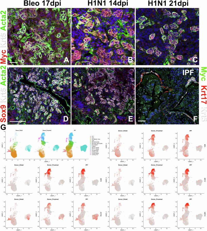
Idiopathic pulmonary fibrosis (IPF) is a progressive respiratory scarring disease arising from the maladaptive differentiation of lung stem cells into bronchial epithelial cells rather than into alveolar type 1 (AT1) cells, which are responsible for gas exchange. Here, we report that healthy lungs maintain their stem cells through tonic Hippo and β-catenin signaling, which promote Yap/Taz degradation and allow for low-level expression of the Wnt target gene Myc. Inactivation of upstream activators of the Hippo pathway in lung stem cells inhibits this tonic β-catenin signaling and Myc expression and promotes their Taz-mediated differentiation into AT1 cells. Vice versa, increased Myc in collaboration with Yap promotes the differentiation of lung stem cells along the basal and myoepithelial-like lineages allowing them to invade and bronchiolize the lung parenchyma in a process reminiscent of submucosal gland development. Our findings indicate that stem cells exhibiting the highest Myc levels become supercompetitors that drive remodeling, whereas loser cells with lower Myc levels terminally differentiate into AT1 cells.

摘要

特发性肺纤维化(IPF)是一种进行性的呼吸性瘢痕疾病,它源于肺干细胞向支气管上皮细胞而非负责气体交换的1型肺泡(AT1)细胞的适应性分化异常。在此,我们报告健康的肺通过持续性的Hippo和β-连环蛋白信号通路维持其干细胞,这两种信号通路促进Yap/Taz降解,并允许Wnt靶基因Myc的低水平表达。肺干细胞中Hippo通路上游激活因子的失活会抑制这种持续性的β-连环蛋白信号传导和Myc表达,并促进它们通过Taz介导分化为AT1细胞。反之,Myc与Yap协同增加,会促进肺干细胞沿基底和肌上皮样谱系分化,使它们能够在类似于黏膜下腺发育的过程中侵入肺实质并使其细支气管化。我们的研究结果表明,表现出最高Myc水平的干细胞成为驱动重塑的超级竞争者,而Myc水平较低的“失败者”细胞则终末分化为AT1细胞。

https://cdn.ncbi.nlm.nih.gov/pmc/blobs/fefb/11621346/e5c09eba6fc4/41467_2024_54997_Fig5_HTML.jpg
https://cdn.ncbi.nlm.nih.gov/pmc/blobs/fefb/11621346/b235431e5a63/41467_2024_54997_Fig1_HTML.jpg
https://cdn.ncbi.nlm.nih.gov/pmc/blobs/fefb/11621346/b644c55cb0b3/41467_2024_54997_Fig2_HTML.jpg
https://cdn.ncbi.nlm.nih.gov/pmc/blobs/fefb/11621346/e5c09eba6fc4/41467_2024_54997_Fig5_HTML.jpg
https://cdn.ncbi.nlm.nih.gov/pmc/blobs/fefb/11621346/b235431e5a63/41467_2024_54997_Fig1_HTML.jpg
https://cdn.ncbi.nlm.nih.gov/pmc/blobs/fefb/11621346/b644c55cb0b3/41467_2024_54997_Fig2_HTML.jpg
https://cdn.ncbi.nlm.nih.gov/pmc/blobs/fefb/11621346/e5c09eba6fc4/41467_2024_54997_Fig5_HTML.jpg

相似文献

1
Cell competition drives bronchiolization and pulmonary fibrosis.细胞竞争驱动细支气管化生和肺纤维化。
Nat Commun. 2024 Dec 5;15(1):10624. doi: 10.1038/s41467-024-54997-2.
2
Cell competition drives bronchiolization and pulmonary fibrosis.细胞竞争驱动细支气管化生和肺纤维化。
Res Sq. 2024 Apr 22:rs.3.rs-4177351. doi: 10.21203/rs.3.rs-4177351/v1.
3
Hippo signaling impairs alveolar epithelial regeneration in pulmonary fibrosis.Hippo 信号通路在肺纤维化中损害肺泡上皮细胞再生。
Elife. 2023 May 11;12:e85092. doi: 10.7554/eLife.85092.
4
YAP/TAZ Signaling in the Pathobiology of Pulmonary Fibrosis.YAP/TAZ 信号通路在肺纤维化发病机制中的作用。
Cells. 2024 Sep 10;13(18):1519. doi: 10.3390/cells13181519.
5
YAP Alleviates Pulmonary Fibrosis Through Promoting Alveolar Regeneration via Modulating the Stemness of Alveolar Type 2 Cells.YAP 通过调节肺泡 II 型细胞的干性来促进肺泡再生,从而减轻肺纤维化。
Stem Cells Dev. 2024 Nov;33(21-22):586-594. doi: 10.1089/scd.2024.0101. Epub 2024 Aug 23.
6
Single-cell transcriptome analysis revealing mechanotransduction via the Hippo/YAP pathway in promoting fibroblast-to-myofibroblast transition and idiopathic pulmonary fibrosis development.单细胞转录组分析揭示通过Hippo/YAP信号通路进行的机械转导在促进成纤维细胞向肌成纤维细胞转变及特发性肺纤维化发展中的作用。
Gene. 2025 Apr 5;943:149271. doi: 10.1016/j.gene.2025.149271. Epub 2025 Jan 22.
7
Role of Hippo-YAP/TAZ signaling pathway in organ fibrosis.河马 - YAP/TAZ信号通路在器官纤维化中的作用。
Zhong Nan Da Xue Xue Bao Yi Xue Ban. 2024;49(9):1509-1516. doi: 10.11817/j.issn.1672-7347.2024.230577.
8
Mechanosignaling through YAP and TAZ drives fibroblast activation and fibrosis.通过YAP和TAZ的机械信号传导驱动成纤维细胞活化和纤维化。
Am J Physiol Lung Cell Mol Physiol. 2015 Feb 15;308(4):L344-57. doi: 10.1152/ajplung.00300.2014. Epub 2014 Dec 12.
9
Role of the Hippo-YAP/TAZ Pathway in Epithelioid Hemangioendothelioma and its Potential as a Therapeutic Target.Hippo-YAP/TAZ 通路在上皮样血管内皮细胞瘤中的作用及其作为治疗靶点的潜力。
Anticancer Res. 2024 Oct;44(10):4147-4153. doi: 10.21873/anticanres.17245.
10
YAP/TAZ activation mediates PQ-induced lung fibrosis by sustaining senescent pulmonary epithelial cells.YAP/TAZ 的激活通过维持衰老的肺上皮细胞介导 PQ 诱导的肺纤维化。
Respir Res. 2024 May 18;25(1):212. doi: 10.1186/s12931-024-02832-z.

引用本文的文献

1
Inhibition of epithelial cell YAP-TEAD/LOX signaling attenuates pulmonary fibrosis in preclinical models.在临床前模型中,抑制上皮细胞YAP-TEAD/LOX信号通路可减轻肺纤维化。
Nat Commun. 2025 Aug 2;16(1):7099. doi: 10.1038/s41467-025-61795-x.
2
Mechanisms and markers of lung ageing in health and disease.健康与疾病状态下肺衰老的机制与标志物
Eur Respir Rev. 2025 Jul 23;34(177). doi: 10.1183/16000617.0233-2024. Print 2025 Jul.
3
Mechanobiology in Action: Biomaterials, Devices, and the Cellular Machinery of Force Sensing.生物力学在行动:生物材料、装置与力传感的细胞机制

本文引用的文献

1
Generation of human alveolar epithelial type I cells from pluripotent stem cells.从多能干细胞生成人肺泡I型上皮细胞。
Cell Stem Cell. 2024 May 2;31(5):657-675.e8. doi: 10.1016/j.stem.2024.03.017. Epub 2024 Apr 19.
2
Epithelial plasticity and innate immune activation promote lung tissue remodeling following respiratory viral infection.上皮细胞可塑性和固有免疫激活促进呼吸道病毒感染后肺组织重塑。
Nat Commun. 2023 Sep 19;14(1):5814. doi: 10.1038/s41467-023-41387-3.
3
Hippo signaling impairs alveolar epithelial regeneration in pulmonary fibrosis.
Biomolecules. 2025 Jun 10;15(6):848. doi: 10.3390/biom15060848.
4
Epithelial-mesenchymal cell competition coordinates fate transitions across tissue compartments during lung development and fibrosis.上皮-间充质细胞竞争在肺发育和纤维化过程中协调跨组织区室的命运转变。
Res Sq. 2025 May 2:rs.3.rs-6189965. doi: 10.21203/rs.3.rs-6189965/v1.
Hippo 信号通路在肺纤维化中损害肺泡上皮细胞再生。
Elife. 2023 May 11;12:e85092. doi: 10.7554/eLife.85092.
4
SARS-CoV-2 replication in airway epithelia requires motile cilia and microvillar reprogramming.SARS-CoV-2 在气道上皮细胞中的复制需要纤毛的运动和微绒毛的重编程。
Cell. 2023 Jan 5;186(1):112-130.e20. doi: 10.1016/j.cell.2022.11.030. Epub 2022 Dec 2.
5
Niche-mediated repair of airways is directed in an occupant-dependent manner.小生境介导的气道修复是一种受居住者影响的定向修复方式。
Cell Rep. 2022 Dec 20;41(12):111863. doi: 10.1016/j.celrep.2022.111863.
6
ΔNp63 drives dysplastic alveolar remodeling and restricts epithelial plasticity upon severe lung injury.ΔNp63驱动发育异常的肺泡重塑,并在严重肺损伤时限制上皮可塑性。
Cell Rep. 2022 Dec 13;41(11):111805. doi: 10.1016/j.celrep.2022.111805.
7
Intermediary Role of Lung Alveolar Type 1 Cells in Epithelial Repair upon Sendai Virus Infection.肺泡 I 型细胞在仙台病毒感染后上皮修复中的中介作用。
Am J Respir Cell Mol Biol. 2022 Sep;67(3):389-401. doi: 10.1165/rcmb.2021-0421OC.
8
The Field of Cell Competition Comes of Age: Semantics and Technological Synergy.细胞竞争领域走向成熟:语义学与技术协同效应
Front Cell Dev Biol. 2022 May 11;10:891569. doi: 10.3389/fcell.2022.891569. eCollection 2022.
9
Somatic Mutation: What Shapes the Mutational Landscape of Normal Epithelia?体细胞突变:正常上皮细胞的突变景观由什么塑造?
Cancer Discov. 2022 Jul 6;12(7):1642-1655. doi: 10.1158/2159-8290.CD-22-0145.
10
Human distal lung maps and lineage hierarchies reveal a bipotent progenitor.人类远端肺图谱和谱系层次结构揭示了一个双能祖细胞。
Nature. 2022 Apr;604(7904):111-119. doi: 10.1038/s41586-022-04541-3. Epub 2022 Mar 30.