Kaneta Hiroki, Shoji Takeshi, Kato Yuichi, Shozen Hideki, Ueki Shinichi, Morita Hiroyuki, Kozuma Yosuke, Adachi Nobuo
Department of Orthopaedic Surgery, Graduate School of Biomedical and Health Sciences, Hiroshima University, Hiroshima, Japan.
Department of Artificial Joints and Biomaterials, Graduate School of Biomedical and Health Sciences, Hiroshima University, Hiroshima, Japan.
Cartilage. 2024 Dec 9:19476035241302978. doi: 10.1177/19476035241302978.
This study aimed to investigate the relationship between clinical findings and the trabecular microstructure of the subchondral bone in patients with hip osteoarthritis (OA) due to developmental dysplasia of the hip (DDH) using multidetector row computed tomography (MDCT).
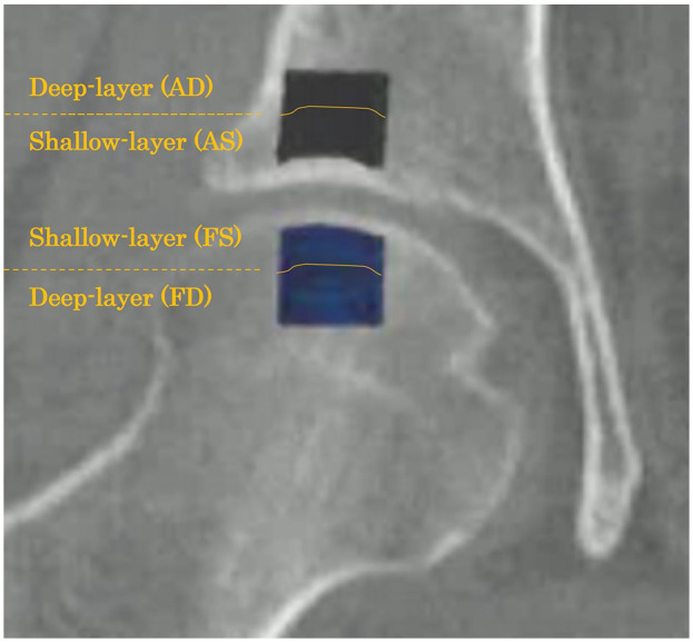
A total of 63 patients (69 hips) with OA due to DDH were retrospectively reviewed, with 12 healthy controls being included for comparison. Clinical evaluation was performed using the Japanese Orthopaedic Association Hip Disease Evaluation Questionnaire (JHEQ). The trabecular bone microstructure was analyzed using MDCT. Regions of interest in the subchondral trabecular bones of the acetabulum and femoral head were defined in the coronal view, and various trabecular microstructural parameters were evaluated.
Bone volume fraction (BV/TV) and trabecular thickness (Tb.Th) exhibited a significant positive correlation with the OA stage, whereas trabecular separation (Tb.Sp) showed a negative correlation. In addition, BV/TV and Tb.Th were negatively correlated with the JHEQ total and pain scores, whereas Tb.Sp was positively correlated with the pain score in all regions.
This is the first study to evaluate the bone microstructure and its relationship with clinical findings in patients with hip OA due to DDH. Our findings suggest that as OA progresses, osteosclerotic changes increase in the acetabulum and femoral head; these changes are associated with worsening clinical symptoms, particularly pain. Targeting the subchondral bone may emerge as a novel treatment strategy for patients with OA due to DDH; nevertheless, further comprehensive studies are required.
本研究旨在利用多排螺旋计算机断层扫描(MDCT)研究发育性髋关节发育不良(DDH)所致髋关节骨关节炎(OA)患者的临床检查结果与软骨下骨小梁微结构之间的关系。
回顾性分析63例(69髋)DDH所致OA患者,并纳入12例健康对照进行比较。采用日本骨科协会髋关节疾病评估问卷(JHEQ)进行临床评估。利用MDCT分析骨小梁微结构。在冠状面上确定髋臼和股骨头软骨下骨小梁的感兴趣区域,并评估各种骨小梁微结构参数。
骨体积分数(BV/TV)和骨小梁厚度(Tb.Th)与OA分期呈显著正相关,而骨小梁间距(Tb.Sp)呈负相关。此外,BV/TV和Tb.Th与JHEQ总分及疼痛评分呈负相关,而Tb.Sp与所有区域的疼痛评分呈正相关。
这是第一项评估DDH所致髋关节OA患者骨微结构及其与临床检查结果关系的研究。我们的研究结果表明,随着OA的进展,髋臼和股骨头的骨硬化改变增加;这些改变与临床症状恶化,尤其是疼痛有关。针对软骨下骨可能成为DDH所致OA患者的一种新的治疗策略;然而,还需要进一步的综合研究。