Center of Experimental Orthopaedics, Saarland University, Kirrberger Straße 100, Building 37, Homburg Saar, D-66421, Germany.
Cartilage Net of the Greater Region, Kirrberger Straße 100, Building 37, Homburg Saar, D-66421, Germany.
Adv Sci (Weinh). 2022 Aug;9(23):e2201692. doi: 10.1002/advs.202201692. Epub 2022 Jun 7.
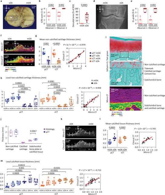
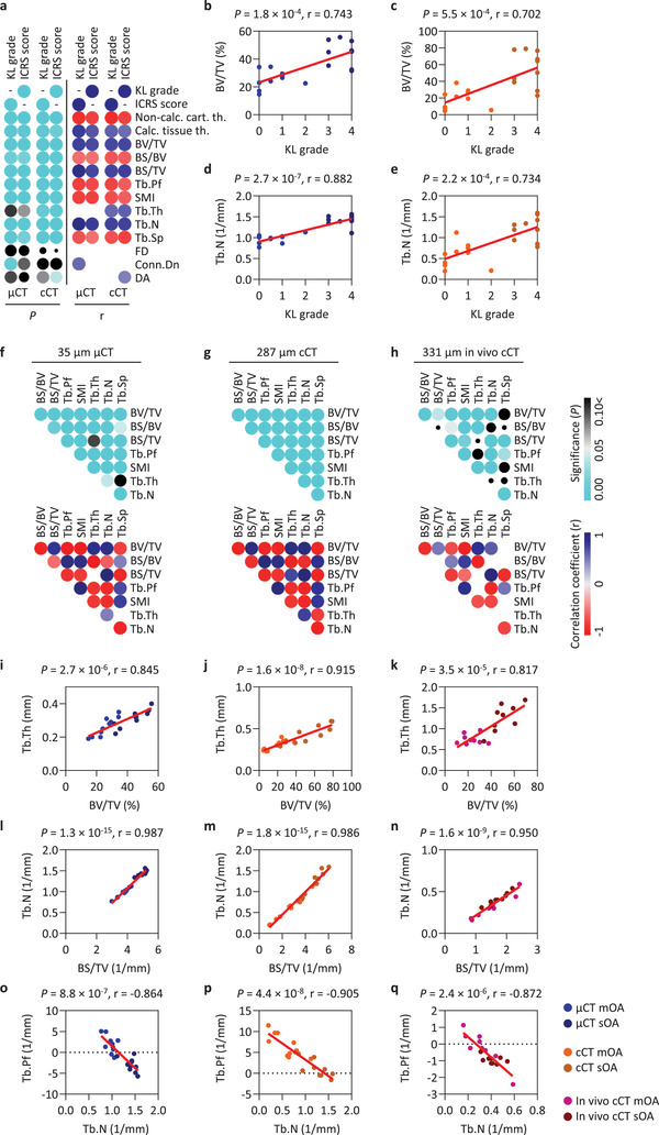
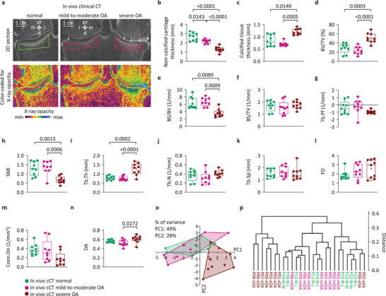
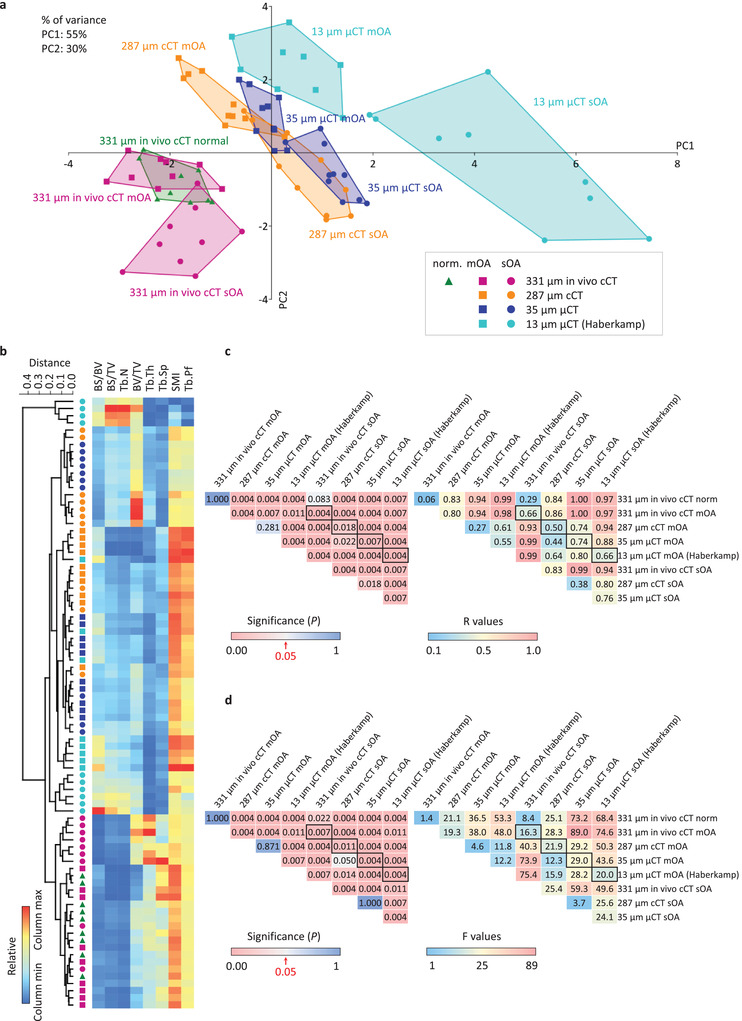
Osteoarthritis (OA) is characterized by critical alterations of the subchondral bone microstructure, besides the well-known cartilaginous changes. Clinical computed tomography (CT) detection of quantitative 3D microstructural subchondral bone parameters is applied to monitor changes of subchondral bone structure in different stages of human OA and is compared with micro-CT, the gold standard. Determination by clinical CT (287 µm resolution) of key microstructural parameters in tibial plateaus with mild-to-moderate and severe OA reveals strong correlations to micro-CT (35 µm), high inter- and intraobserver reliability, and small relative differences. In vivo, normal, mild-to-moderate, and severe OA are compared with clinical CT (331 µm). All approaches detect characteristic expanded trabecular structure in severe OA and fundamental microstructural correlations with clinical OA stage. Multivariate analyses at various in vivo and ex vivo imaging resolutions always reliably separate mild-to-moderate from severe OA (except mild-to-moderate OA from normal), revealing a striking similarity between 287 µm clinical and 35 µm micro-CT. Thus, accurate structural measurements using clinical CT with a resolution near the trabecular dimensions are possible. Clinical CT offers an opportunity to quantitatively monitor subchondral bone microstructure in clinical and experimental settings as an advanced tool of investigating OA and other diseases affecting bone architecture.
骨关节炎(OA)的特征是软骨下骨微观结构的重大改变,除了众所周知的软骨变化。临床计算机断层扫描(CT)检测定量 3D 微观结构软骨下骨参数,用于监测不同阶段人类 OA 软骨下骨结构的变化,并与金标准微 CT 进行比较。使用临床 CT(287µm 分辨率)测定轻度至中度和重度 OA 胫骨平台的关键微观结构参数,与微 CT(35µm)具有很强的相关性,具有很高的观察者内和观察者间可靠性,并且相对差异较小。在体内,将正常、轻度至中度和重度 OA 与临床 CT(331µm)进行比较。所有方法都在严重 OA 中检测到特征性扩张的小梁结构,并与临床 OA 阶段的基本微观结构相关。在不同的体内和离体成像分辨率下进行的多元分析总是可靠地区分轻度至中度和重度 OA(轻度至中度 OA 与正常除外),这表明 287µm 临床 CT 和 35µm 微 CT 之间存在惊人的相似性。因此,使用接近小梁尺寸的临床 CT 进行准确的结构测量是可能的。临床 CT 为在临床和实验环境中定量监测软骨下骨微观结构提供了机会,是研究 OA 和其他影响骨结构疾病的先进工具。