Merbl Yael, Kaur Sukhmeen, Kei Tiffany G, Ryan Elle, Johnson Philippa J
Department of Clinical Sciences, College of Veterinary Medicine, Cornell University, Ithaca, NY, United States.
Front Vet Sci. 2024 Nov 26;11:1434447. doi: 10.3389/fvets.2024.1434447. eCollection 2024.
Describe and characterize the magnetic resonance imaging (MRI) appearance of annulus fibrosus (AF) high-intensity zone (HIZ) in dogs suffering from intervertebral disc disease (IVDD).
A single-center retrospective case series study. Databases were reviewed from 2011 to 2022 for dogs that underwent MRI diagnosis due to suspected IVDD. Cases were included if they had T2-weighted (T2W) hyperintense annular fibrosus lesions (AFL) on the imaging diagnosis report. To be included, the MRI scan had to be of diagnostic quality and include a sagittal T2W, proton density (PD), or short tau inversion recovery (STIR) sequence of the annular lesion, together with transverse T2W and/or dorsal plane STIR sequences over the HIZ region.
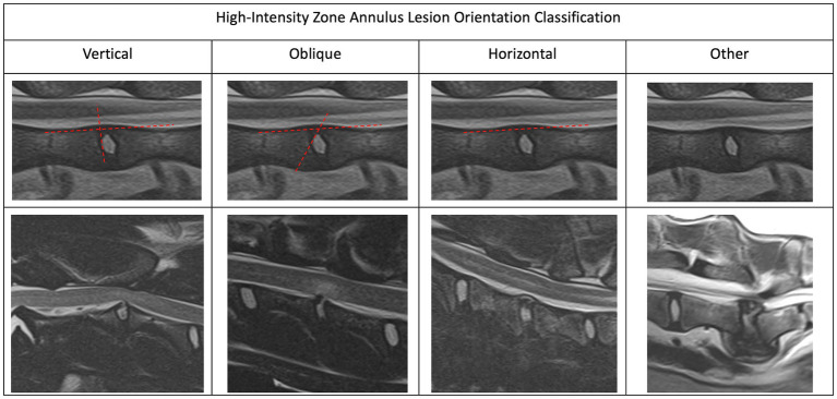
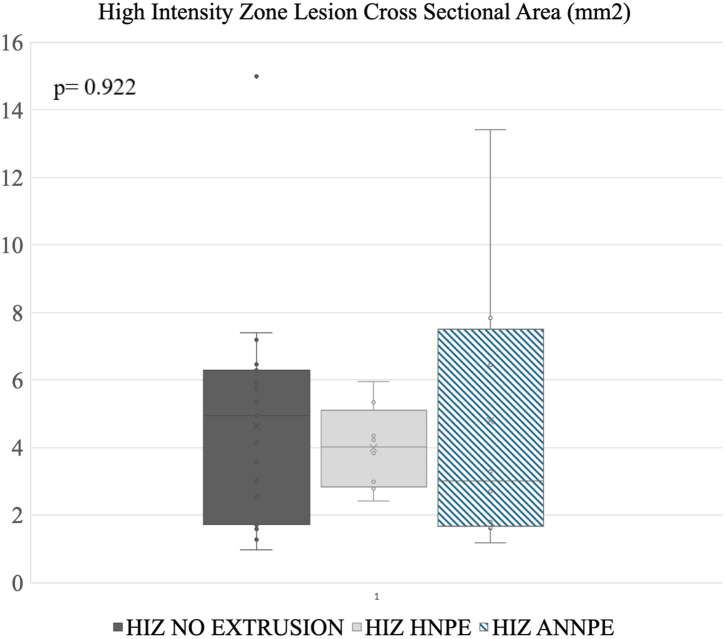
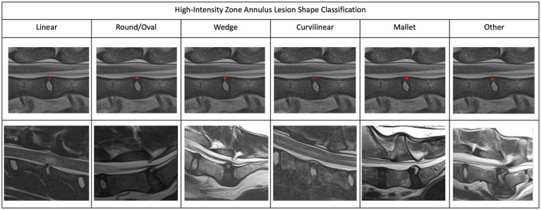
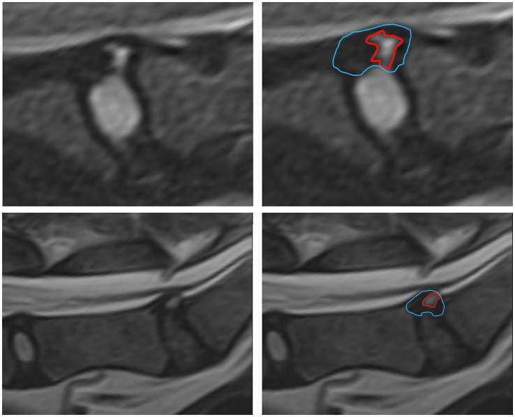
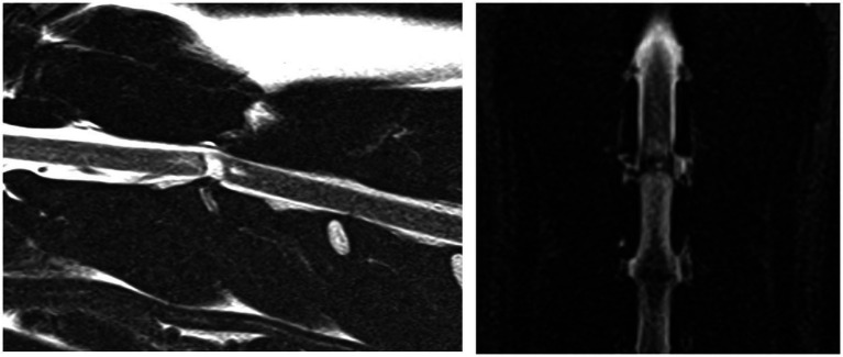
Forty one cases (in 39 dogs) of HIZ were included in the study. Mixed breed dogs were the highest represented group representing 25.6% of the cohort. Patient median age was 7.5 years and median weight 23 kg. Primary HIZ appeared in 7/39 dogs (17.95%) and the remaining had acute non-compressive nucleus pulposus extrusion (ANNPE), hydrated nucleus pulposus extrusion (HNPE) or concurrent myelopathy. Characterization of HIZ lesions included several variable appearances in orientation and shape. HIZ lesions were most easily identifiable in the sagittal plane. Similar to humans, the most common site of HIZ without extrusion was the lumbosacral (LS) region. All the dogs with HIZ lesions as the most significant MRI finding, exhibited spinal pain and/or chronic paresis/plegia.
By introducing and defining HIZ lesions to the veterinary imaging nomenclature, we hope future studies will further examine the prevalence and clinical significance of HIZ lesions in canine patients.
描述和表征患有椎间盘疾病(IVDD)的犬类纤维环(AF)高强度区(HIZ)的磁共振成像(MRI)表现。
一项单中心回顾性病例系列研究。对2011年至2022年因疑似IVDD接受MRI诊断的犬类数据库进行了回顾。如果成像诊断报告中有T2加权(T2W)高信号的纤维环病变(AFL),则纳入病例。要纳入研究,MRI扫描必须具有诊断质量,并且包括环形病变的矢状面T2W、质子密度(PD)或短tau反转恢复(STIR)序列,以及HIZ区域的横断面T2W和/或背侧平面STIR序列。
该研究纳入了41例(39只犬)HIZ病例。混血犬是占比最高的群体,占队列的25.6%。患者中位年龄为7.5岁,中位体重为23千克。原发性HIZ出现在7/39只犬(17.95%)中,其余犬患有急性非压迫性髓核突出(ANNPE)、水合髓核突出(HNPE)或并发脊髓病。HIZ病变的特征包括方向和形状的几种可变表现。HIZ病变在矢状面最容易识别。与人类相似,无突出的HIZ最常见部位是腰骶(LS)区域。所有以HIZ病变为最重要MRI表现的犬均表现出脊柱疼痛和/或慢性轻瘫/瘫痪。
通过将HIZ病变引入并定义到兽医影像学命名中,我们希望未来的研究将进一步检查HIZ病变在犬类患者中的患病率和临床意义。