• 文献检索
  • 文档翻译
  • 深度研究
  • 学术资讯
  • Suppr Zotero 插件Zotero 插件
  • 邀请有礼
  • 套餐&价格
  • 历史记录
应用&插件
Suppr Zotero 插件Zotero 插件浏览器插件Mac 客户端Windows 客户端微信小程序
定价
高级版会员购买积分包购买API积分包
服务
文献检索文档翻译深度研究API 文档MCP 服务
关于我们
关于 Suppr公司介绍联系我们用户协议隐私条款
关注我们

Suppr 超能文献

核心技术专利:CN118964589B侵权必究
粤ICP备2023148730 号-1Suppr @ 2026

文献检索

告别复杂PubMed语法,用中文像聊天一样搜索,搜遍4000万医学文献。AI智能推荐,让科研检索更轻松。

立即免费搜索

文件翻译

保留排版,准确专业,支持PDF/Word/PPT等文件格式,支持 12+语言互译。

免费翻译文档

深度研究

AI帮你快速写综述,25分钟生成高质量综述,智能提取关键信息,辅助科研写作。

立即免费体验

构建用于估计全血和脑肿瘤血管中R及R*弛豫与钆喷酸葡胺浓度关系的逼真三维模型之路

The Road to a Realistic 3D Model for Estimating R and R* Relaxation Versus Gd-DTPA Concentration in Whole Blood and Brain Tumor Vasculature.

作者信息

van Dorth Daniëlle, Alafandi Ahmad, Soloukey Sadaf, Kruizinga Pieter, Venugopal Krishnapriya, Delphin Aurélien, Poot Dirk H J, Christen Thomas, Smits Marion, de Bresser Jeroen, Hernandez-Tamames Juan A, van Osch Matthias J P

机构信息

C. J. Gorter MRI Center, Department of Radiology, Leiden University Medical Center, Leiden, The Netherlands.

Department of Radiology and Nuclear Medicine, Erasmus MC, University Medical Center Rotterdam, Rotterdam, The Netherlands.

出版信息

NMR Biomed. 2025 Jan;38(1):e5308. doi: 10.1002/nbm.5308.

DOI:10.1002/nbm.5308
PMID:39663686
原文链接:https://pmc.ncbi.nlm.nih.gov/articles/PMC11635185/
Abstract

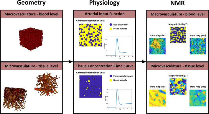
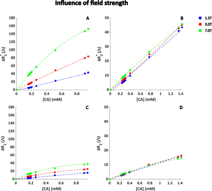
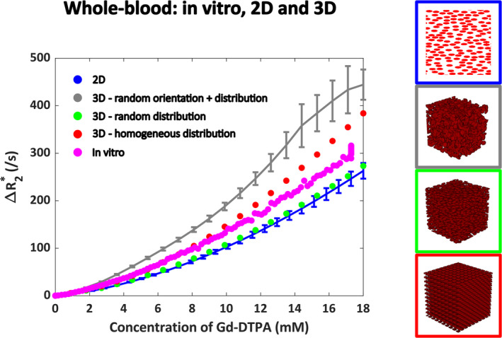
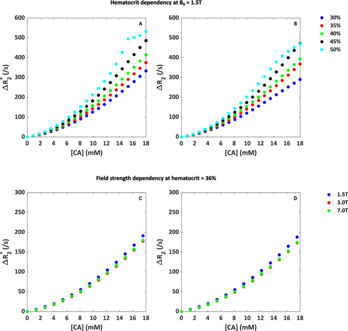
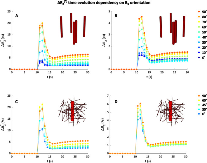
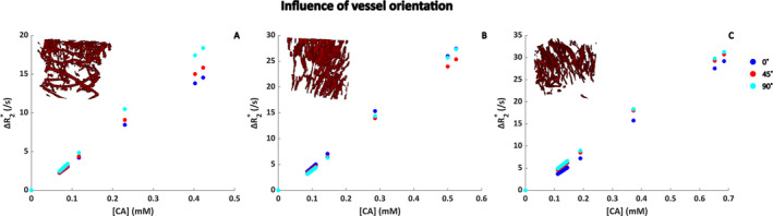
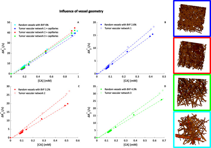
Dynamic susceptibility contrast (DSC) MRI is commonly part of brain tumor imaging. For quantitative analysis, measurement of the arterial input function and tissue concentration time curve is required. Usually, a linear relationship between the MR signal changes and contrast agent concentration ([Gd]) is assumed, even though this is a known simplification. The aim of this study was to develop a realistic 3D simulation model as an efficient method to assess the relationship between ΔR and [Gd] both in whole blood and brain tissue. We modified an open-source 3D simulation model to study different red blood cell configurations for assessing whole-blood ΔR versus [Gd]. The results were validated against previously obtained 2D data and in vitro data. Furthermore, hematocrit levels (30%-50%) and field strengths (1.5-3.0-7.0 T) were varied. Subsequently, realistic tumor vascular networks were derived from intraoperative high framerate Doppler ultrasound data to study the influence of vascular structure and orientation with respect to the main magnetic field (1.5-3.0-7.0 T) for the calculation of ΔR versus [Gd] in brain tissue. For whole blood, good agreement of the 3D model was found with in vitro and 2D simulation data when red blood cells were aligned with the blood flow. For brain tissue, minor differences were found between the vascular networks. The effect of vessel direction with respect to B was apparent in case of clear directionality of the main vessels. The dependency on field strength agreed with previous reports. In conclusion, we have shown that the relationship between ΔR and [Gd] is affected by the organization of red blood cells and orientation of blood vessels with respect to the main magnetic field, as well as the field strength. These findings are important for further optimization of the realistic 3D model that could eventually be used to improve the estimation of hemodynamic parameters from DSC-MRI.

摘要

动态磁敏感对比(DSC)磁共振成像通常是脑肿瘤成像的一部分。对于定量分析,需要测量动脉输入函数和组织浓度-时间曲线。通常,尽管这是一种已知的简化方法,但仍假定磁共振信号变化与造影剂浓度([Gd])之间存在线性关系。本研究的目的是开发一个逼真的三维模拟模型,作为评估全血和脑组织中ΔR与[Gd]之间关系的有效方法。我们修改了一个开源三维模拟模型,以研究不同红细胞配置对评估全血ΔR与[Gd]的影响。结果与先前获得的二维数据和体外数据进行了验证。此外,还改变了血细胞比容水平(30%-50%)和场强(1.5-3.0-7.0 T)。随后,从术中高帧率多普勒超声数据中得出逼真的肿瘤血管网络,以研究血管结构和相对于主磁场(1.5-3.0-7.0 T)的方向对脑组织中ΔR与[Gd]计算的影响。对于全血,当红细胞与血流方向一致时,三维模型与体外和二维模拟数据具有良好的一致性。对于脑组织,在血管网络之间发现了微小差异。在主血管具有明显方向性的情况下,血管方向相对于B的影响是明显的。对场强的依赖性与先前的报道一致。总之,我们已经表明,ΔR与[Gd]之间的关系受红细胞的组织、血管相对于主磁场的方向以及场强的影响。这些发现对于进一步优化逼真的三维模型很重要,该模型最终可用于改善从DSC-MRI估计血流动力学参数。

https://cdn.ncbi.nlm.nih.gov/pmc/blobs/edf4/11635185/9b682df810f4/NBM-38-e5308-g005.jpg
https://cdn.ncbi.nlm.nih.gov/pmc/blobs/edf4/11635185/a83730a35ed1/NBM-38-e5308-g008.jpg
https://cdn.ncbi.nlm.nih.gov/pmc/blobs/edf4/11635185/393e01b52016/NBM-38-e5308-g002.jpg
https://cdn.ncbi.nlm.nih.gov/pmc/blobs/edf4/11635185/bc6d692d8d10/NBM-38-e5308-g003.jpg
https://cdn.ncbi.nlm.nih.gov/pmc/blobs/edf4/11635185/70f119392395/NBM-38-e5308-g006.jpg
https://cdn.ncbi.nlm.nih.gov/pmc/blobs/edf4/11635185/28b793de2989/NBM-38-e5308-g007.jpg
https://cdn.ncbi.nlm.nih.gov/pmc/blobs/edf4/11635185/10c9ce76802e/NBM-38-e5308-g001.jpg
https://cdn.ncbi.nlm.nih.gov/pmc/blobs/edf4/11635185/9b682df810f4/NBM-38-e5308-g005.jpg

相似文献

1
The Road to a Realistic 3D Model for Estimating R and R* Relaxation Versus Gd-DTPA Concentration in Whole Blood and Brain Tumor Vasculature.构建用于估计全血和脑肿瘤血管中R及R*弛豫与钆喷酸葡胺浓度关系的逼真三维模型之路
NMR Biomed. 2025 Jan;38(1):e5308. doi: 10.1002/nbm.5308.
2
Dependency of R and R * relaxation on Gd-DTPA concentration in arterial blood: Influence of hematocrit and magnetic field strength.动脉血中 Gd-DTPA 浓度对 R 和 R * 弛豫的依赖性:血细胞比容和磁场强度的影响。
NMR Biomed. 2022 May;35(5):e4653. doi: 10.1002/nbm.4653. Epub 2021 Nov 23.
3
Intraindividual in vivo comparison of gadolinium contrast agents for pharmacokinetic analysis using dynamic contrast enhanced magnetic resonance imaging.应用动态对比增强磁共振成像进行药代动力学分析的钆对比剂的个体内体内比较。
Invest Radiol. 2010 May;45(5):233-44. doi: 10.1097/RLI.0b013e3181d54507.
4
Intra-individual comparison of different gadolinium-based contrast agents in the quantitative evaluation of C6 glioma with dynamic contrast-enhanced magnetic resonance imaging.动态对比增强磁共振成像定量评估 C6 胶质瘤中不同钆基对比剂的个体内比较。
Sci China Life Sci. 2017 Jan;60(1):11-15. doi: 10.1007/s11427-016-0386-2. Epub 2017 Jan 4.
5
Correlation of Tumor Perfusion Between Carbon-13 Imaging with Hyperpolarized Pyruvate and Dynamic Susceptibility Contrast MRI in Pre-Clinical Model of Glioblastoma.碳-13 成像与动态对比磁共振成像在神经胶质瘤临床前模型中对肿瘤灌注的相关性研究
Mol Imaging Biol. 2019 Aug;21(4):626-632. doi: 10.1007/s11307-018-1275-y.
6
Consistency of normalized cerebral blood volume values in glioblastoma using different leakage correction algorithms on dynamic susceptibility contrast magnetic resonance imaging data without and with preload.动磁敏感对比磁共振成像数据在无和有预加载情况下,使用不同漏出校正算法对胶质母细胞瘤标准化脑血容量值的一致性。
J Neuroradiol. 2019 Feb;46(1):44-51. doi: 10.1016/j.neurad.2018.04.006. Epub 2018 May 21.
7
Approximation of arterial input curve data in MRI estimation of cerebral blood-tumor-barrier leakage: comparison between Gd-DTPA and 99mTc-DTPA input curves.MRI评估脑血肿瘤屏障渗漏时动脉输入曲线数据的近似:钆喷酸葡胺与锝[99mTc]喷替酸输入曲线的比较
Magn Reson Imaging. 1996;14(3):235-41. doi: 10.1016/0730-725x(95)02082-5.
8
Assessment of the effect of haematocrit-dependent arterial input functions on the accuracy of pharmacokinetic parameters in dynamic contrast-enhanced MRI.评估血球比容依赖性动脉输入函数对动态对比增强 MRI 中药代动力学参数准确性的影响。
NMR Biomed. 2011 Aug;24(7):902-15. doi: 10.1002/nbm.1648. Epub 2011 Feb 3.
9
Pharmacokinetic approach for dynamic breast MRI to indicate signal intensity time curves of benign and malignant lesions by using the tumor flow residence time.利用肿瘤血流滞留时间对动态乳腺 MRI 的药代动力学方法,对良、恶性病变的信号强度时间曲线进行指示。
Invest Radiol. 2013 Feb;48(2):69-78. doi: 10.1097/RLI.0b013e31827d29cf.
10
Multiparametric characterization of response to anti-angiogenic therapy using USPIO contrast-enhanced MRI in combination with dynamic contrast-enhanced MRI.使用超顺磁氧化铁对比增强 MRI 联合动态对比增强 MRI 进行多参数特征分析以评估抗血管生成治疗的反应。
J Magn Reson Imaging. 2018 Jun;47(6):1589-1600. doi: 10.1002/jmri.25898. Epub 2017 Dec 4.

本文引用的文献

1
Human brain mapping using co-registered fUS, fMRI and ESM during awake brain surgeries: A proof-of-concept study.在清醒脑手术中使用配准的 fUS、fMRI 和 ESM 进行人脑绘图:概念验证研究。
Neuroimage. 2023 Dec 1;283:120435. doi: 10.1016/j.neuroimage.2023.120435. Epub 2023 Oct 30.
2
The role of DSC MR perfusion in predicting IDH mutation and 1p19q codeletion status in gliomas: meta-analysis and technical considerations.DSC MR 灌注在预测胶质瘤 IDH 突变和 1p19q 共缺失状态中的作用:荟萃分析和技术考虑。
Neuroradiology. 2023 Jul;65(7):1111-1126. doi: 10.1007/s00234-023-03154-5. Epub 2023 May 13.
3
Microvasculature Features Derived from Hybrid EPI MRI in Non-Enhancing Adult-Type Diffuse Glioma Subtypes.
基于混合回波平面成像磁共振成像的非强化成人型弥漫性胶质瘤亚型的微血管特征
Cancers (Basel). 2023 Apr 4;15(7):2135. doi: 10.3390/cancers15072135.
4
Dependency of R and R * relaxation on Gd-DTPA concentration in arterial blood: Influence of hematocrit and magnetic field strength.动脉血中 Gd-DTPA 浓度对 R 和 R * 弛豫的依赖性:血细胞比容和磁场强度的影响。
NMR Biomed. 2022 May;35(5):e4653. doi: 10.1002/nbm.4653. Epub 2021 Nov 23.
5
Consensus recommendations for a dynamic susceptibility contrast MRI protocol for use in high-grade gliomas.用于高级别脑胶质瘤的动态对比磁共振成像协议的共识建议。
Neuro Oncol. 2020 Sep 29;22(9):1262-1275. doi: 10.1093/neuonc/noaa141.
6
Differentiation of progressive disease from pseudoprogression using 3D PCASL and DSC perfusion MRI in patients with glioblastoma.使用 3D PCASL 和 DSC 灌注 MRI 区分胶质母细胞瘤患者的进行性疾病与假性进展。
J Neurooncol. 2020 May;147(3):681-690. doi: 10.1007/s11060-020-03475-y. Epub 2020 Apr 1.
7
Quantitative theory for the transverse relaxation time of blood water.血液水横向弛豫时间的定量理论。
NMR Biomed. 2020 May;33(5):e4207. doi: 10.1002/nbm.4207. Epub 2020 Feb 5.
8
Functional Ultrasound (fUS) During Awake Brain Surgery: The Clinical Potential of Intra-Operative Functional and Vascular Brain Mapping.清醒开颅手术中的功能超声(fUS):术中脑功能和血管成像的临床潜力
Front Neurosci. 2020 Jan 9;13:1384. doi: 10.3389/fnins.2019.01384. eCollection 2019.
9
Simultaneous R and quantitative susceptibility mapping measurement enables differentiation of thrombus hematocrit and age: an in vitro study at 3 T.同时进行 R 及定量磁化率图测量可区分血栓血细胞比容和年龄:3T 体外研究。
J Neurointerv Surg. 2019 Nov;11(11):1155-1161. doi: 10.1136/neurintsurg-2019-014802. Epub 2019 May 14.
10
Validation of vessel size imaging (VSI) in high-grade human gliomas using magnetic resonance imaging, image-guided biopsies, and quantitative immunohistochemistry.采用磁共振成像、影像引导下活检和定量免疫组织化学技术验证高级别人脑胶质瘤中的血管大小成像(VSI)。
Sci Rep. 2019 Feb 26;9(1):2846. doi: 10.1038/s41598-018-37564-w.